
- Title
-
Analysis of maternal-zygotic ugdh mutants reveals divergent roles for HSPGs in vertebrate embryogenesis and provides new insight into the initiation of left-right asymmetry
- Authors
- Superina, S., Borovina, A., and Ciruna, B.
- Source
- Full text @ Dev. Biol.
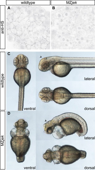
MZjek mutant embryos lack extracellular heparan sulfate proteoglycans and display multiple morphological abnormalities. (A,B) Representative confocal micrographs of anti- heparan sulfate PG staining in the animal pole of shield-staged wildtype (A) and MZjek mutant (B) embryos (N=20,20 respectively). Anti-heparan sulfate staining is clearly present in the extracellular space of wildtype embryos. In MZjek, there is an absence of HS staining, suggesting HS GAGs are not synthesized in the mutant. (C,D) Live images of 48 hpf WT (C) and MZjek mutant embryos (D) mounted for lateral, dorsal and ventral views. MZjek (D) exhibit an expanded mediolateral axis (bars), defects in axial extension, brain patterning defects (arrowheads) and disrupted heart morphogenesis. |

MZjek have a dysmorphic heart and reduced ventricular populations. (A,B) Lateral views of hearts stained with MF20 (red) and S46 (green) antibodies to visualize the ventricle and atrium of 48 hpf embryos. MF20 marks the entire heart and S46 is atrium specific. In these superimposed images MF20+S46- ventricle is red and MF20+S46+ atrium appears yellow. (A) Wildtype heart with two distinct chambers. (B) MZjek display a dysmorphic heart, with a misshapen atrium and a strung out ventricle (N=9). (C–F) amhc and vmhc expression in wildtype (C,E) and MZjek (D,F) at the 20-somite stage. MZjek mutants exhibit reduced expression of vmhc when compared to similarly staged wildtype embryos (N=25, 36 respectively). EXPRESSION / LABELING:
PHENOTYPE:
|
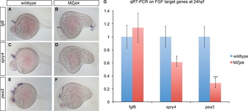
Fgf signaling is reduced in MZjek. (A–F) WISH for fgf8, spry4 and pea3 in wildtype (A,C,E; tailbud out of frame) and MZjek (B,D,F) embryos. In MZjek, fgf8 is present and expressed at equivalent levels to wildtype embryos (A,B; N=62,60), however targets spry4 (C,D; N=92,75) and pea3 (E,F; N=35,39) appear reduced. (G) qRT-PCR assays of fgf8 (p=0.2257), spry4 (*p<0.05), pea3 (**p<0.01) expression in wildtype versus MZjek embryos at 24 hpf. Error bars represent the s.e. for the expression level fold change. |
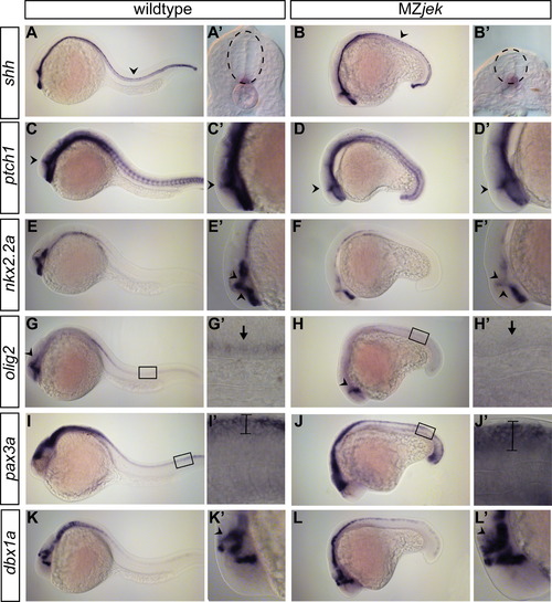
Shh signaling is reduced in MZjek. (A,B) shh is expressed in the notochord and floorplate (arrowhead) of wildtype (A, N=30) and MZjek at 24 hpf (B, N=40). Insets show transverse sections of shh in the spinal cord (A',B' N=28,39). (C–H) Class II genes patched1, nkx2.2a, and olig2 are reduced in MZjek. patched1 expression is reduced in MZjek (D, N=26) as compared to wildtype embryos (C, N=18). Most notably, patched1 expression is severely reduced in the ZLI (arrowheads C,D). Insets show close up of head region, highlighting expression in the ZLI (arrowheads C′,D′). nkx2.2a is absent in prethalamic (anterior arrowhead) and thalamic (posterior arrowhead) areas surrounding the ZLI in MZjek mutants (E,E',F,F' N=25,37). olig2 expression is reduced in the forebrain (G,H arrowhead; N=12,17) and ventral spinal cord (arrow in insets G'H') in MZjek embryos. Boxed region in G,H shown at higher magnification in G′,H′. (I–L) Class I genes pax3a and dbx1a show increased expression in MZjek. pax3a expression is expanded in the dorsal spinal cord (bars) in MZjek embryos (I,I',J,J' N=23,34). Boxed region in I,J shown at higher magnification in I',J'. dbx1a expression is also expanded in the thalamic areas posterior to the ZLI (arrowheads K,K′,L,'' N=8,10). EXPRESSION / LABELING:
|
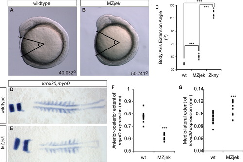
Convergence and extension defects are apparent in MZjek. (A,B) Representative photos of live wildtype (A; N=9) and MZjek (B; N=7) embryos at 9 SS. (C) Quantification of the angle of body axis extension over the yolk in wildtype, MZjek and Zkny (***p<0.001). (D,E) Flat-mounts of 10 SS wildtype (D; N=13) and MZjek (E; N=10) embryos stained for krox-20 (hindbrain) and myoD (somite) gene expression. (F) Quantification of the anterior–posterior extent of the myoD expression domain in wildtype versus MZjek (***p<0.001). (G) Quantification of the mediolateral extent of krox20 expression in wildtype versus MZjek (***p<0.001). EXPRESSION / LABELING:
PHENOTYPE:
|
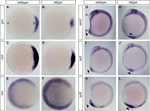
Canonical Wnt signaling is upregulated in MZjek;. (A–F) Whole-mount in situ hybridization (WISH) for dorsal organizer genes goosecoid (A,B; gsc; N=26,23) and chordin (C,D; chd; N=20,21), and ventral gene vox (E,F; N=15,18) at shield stage. Dorsal (D) and Ventral (V) orientation indicated in panel A. Embryos are viewed from the animal pole with dorsal to the right. (G–L) WISH of axin2 (G,H; N=12,8), sp5 (I,J; N=18,11), and sp5l (K,L; N=9,8) at 12 SS. Gene expression appears to be most dramatically upregulated in the tailbud (arrowheads). Anterior (A) and Posterior (P) extents are as indicated in panel G. EXPRESSION / LABELING:
PHENOTYPE:
|

MZjek mutants show left–right patterning defects despite having intact embryonic midlines. (A–D) WISH for lefty1,2 (A,C; N=80,116) and spaw (B,D; N=16,43) in wildtype and MZjek embryos at 21 SS. (E) Percentage of embryos displaying left, right, bilateral and absent lefty1,2 and spaw expression in 20–22 SS wildtype (wt) and MZjek mutant embryos. (F–K) Midline markers shh (F,I), ntl (G,J), and lefty1 (H,K) are expressed continuously along the embryonic axis in both wildtype and MZjek embryos, indicating an intact midline. EXPRESSION / LABELING:
PHENOTYPE:
|
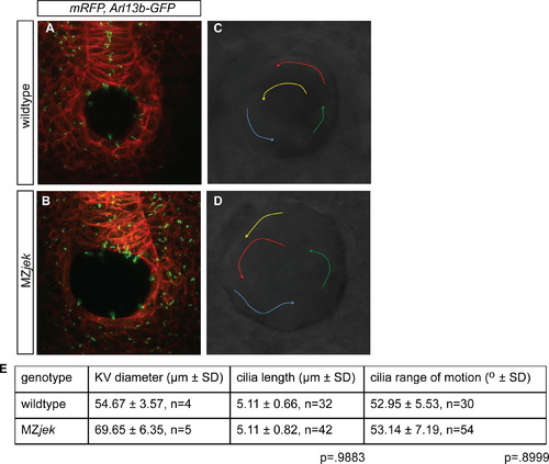
MZjek mutants have normal cilia length and normal fluid flow within Kupfferós vesicle (KV). (A,B) Confocal z-stack images through the KV of 6–8 SS wildtype (A) and MZjek (B) embryos that express membrane-localized mRFP (red) and Arl13b-GPF (green) demonstrate the presence of motile cilia. (C,D) Bead trajectories in wildtype and MZjek KVs injected with fluorescent beads at 8–10 SS. Representative bead trajectories in wildtype (C) and MZjek (D) indicated by arrows. (E) Mean and standard deviation of KV diameter, motile cilia length and range of motion of cilia in the KV of wildtype and MZjek embryos. PHENOTYPE:
|
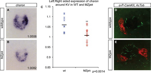
Symmetry breaking in MZjek is abnormal downstream of flow establishment. (A,B) charon expression at KV in wildtype (A) and MZjek (B) embryos at 14 SS with corresponding ratios of left:right side expression quantification. (C) Ratio of left sided:right sided expression of charon surrounding KV in wildtype and MZjek (**p<0.01). (D,E) Confocal immunofluorescence of KV using anti-acetylated alpha-tubulin (green) and anti-phosphorylated CaMK-II (red) to visualize activated CaMK-II in cells surrounding the KV in wildtype (D) and MZjek (E) embryos at 12 SS (N=26, ***p<0.001). EXPRESSION / LABELING:
PHENOTYPE:
|
Reprinted from Developmental Biology, 387(2), Superina, S., Borovina, A., and Ciruna, B., Analysis of maternal-zygotic ugdh mutants reveals divergent roles for HSPGs in vertebrate embryogenesis and provides new insight into the initiation of left-right asymmetry, 154-166, Copyright (2014) with permission from Elsevier. Full text @ Dev. Biol.