
- Title
-
The cdx Genes and Retinoic Acid Control the Positioning and Segmentation of the Zebrafish Pronephros
- Authors
- Wingert, R.A., Selleck, R., Yu, J., Song, H.D., Chen, Z., Song, A., Zhou, Y., Thisse, B., Thisse, C., McMahon, A.P., and Davidson, A.J.
- Source
- Full text @ PLoS Genet.

The Zebrafish Pronephric Nephrons Consist of Multiple Tubule Segments Similar to Metanephric Nephrons. (A) Whole-mount in situ hybridization of wild-type embryos shows segmentation of each pronephric nephron into podocytes (Pod), neck (N), proximal convoluted tubule (PCT), proximal straight tubule (PST), distal early (DE), corpuscle of Stannius (CS), distal late (DL), and pronephric duct (PD) that fuse to the cloaca (C) (not shown). Lateral views are shown except for dorsal view in top left panel, with anterior to the left. (B) Murine section in situ hybridzation of solute transporters, bar denotes 500 μm. (C) Time course of PCT convolution and adjacent PST, with slc20a1a expression used to label the PCT at all stages, PST labeled by trpm7 expression at 26 hpf, and slc13a1 expression at 48?144 hpf. (D) Expression of cdh17, slc9a3, and rfx2 at 48 hpf in distinct segments of the pronephros. Note expression of rfx2 in the presumptive ciliated neck segment. (C and D) Dorsal views are shown, with anterior to the left. (E) Schema of the original model of the 48 hpf zebrafish pronephros (top), our redefined model showing segmentation of each nephrons (middle), and a mammalian nephron stretched out to compare segmentation of the metanephric nephron (bottom). Mammalian segments are color coded according to observations of shared solute transporter expressions with respective zebrafish pronephros segments. Additional abbreviations are: G (glomerulus), N (neck), TL (thin limb), TAL (thick ascending limb), MD (macula densa), DCT (distal convoluted tubule), CNT (connecting tubule), and CD (collecting duct). |
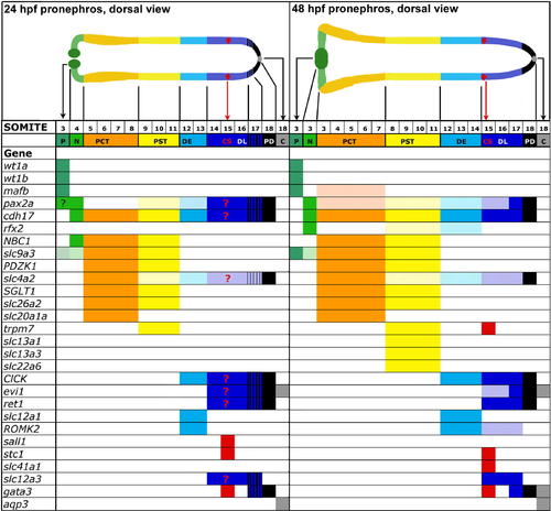
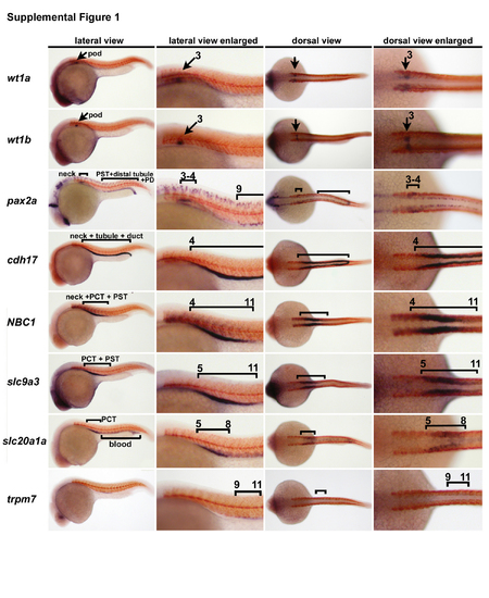
Pronephros Expression of Known Renal Genes and Solute Transporters Isolated Using Functional Genomic. The domain of each pronephros-expressed gene was mapped along the embryonic axis according to somite boundaries, using double in situ hybridization with mhc to label the myotomes. Solid color bars indicate strong expression, light color bars indicate weak expression, and blue/black stripe bars indicate overlap in the expression domains of DL and PD markers; note that rfx2 expression at 48 hpf is discontinuous in the PST and DE domains. Schematic anatomy of the pronephros at 24 hpf (top, left) compared to 48 hpf (top, right). The cloaca (C) is not considered a segment of the pronephros, but is shown to depict the terminus of the pronephros. |

RA Signaling Is Required for Proximal Segment Fates and to Prevent the Expansion of Distal Segment Fates. (A) Expression of segment markers and mhc at 48 hpf in wild-type embryos and raldh2 morphants, and wild-type embryos that were incubated with either DMSO (control) or DEAB for indicated times during development, by double whole-mount in situ hybridization. Brackets and arrows indicate expression domains. Abbreviations are: %e (percent epiboly), s (somite stage). Dorsal views are shown, with anterior to the left, with the exception of lateral views for gata3 and aqp3 expression. (B) Summary of nephron segmentation with respect to embryo somite number for each DEAB treatment. |
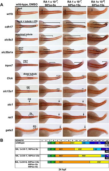
Exogenous RA Treatment Proximalizes the Pronephros. (A) Expression of segment markers and mhc at 24 hpf in wild-type embryos incubated with either DMSO (control) or RA at the indicated dosage and times during development, by double whole-mount in situ hybridization. Brackets and arrows indicate expression domains. Abbreviations are: %e (percent epiboly), s (somite stage). Dorsal views are shown, with anterior to the left, with the exception of lateral views for gata3 expression. (B) Summary of nephron segmentation with respect to embryo somite number for each RA experiment. |
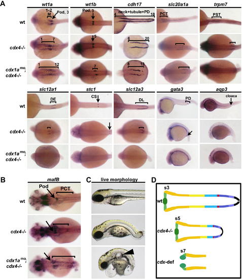
cdx Genes Position the Pronephros along the Embryonic Axis and Are Requisite for Distal Segment Formation. Expression of segment markers was performed on wild-type, cdx4?/? mutants, and cdx-deficient embryos at (A) 26 hpf and (B) 48 hpf using whole-mount in situ hybridization. Brackets and arrows indicate expression domains in each embryo. Numbers indicate somite number. Dorsal views of embryos, with anterior to the left, with the exception of lateral views for gata3 and aqp3 expression. (C) Live wild-type, cdx4?/? mutants, and cdx-deficient embryos at 72 hpf. Arrowhead indicates glomerular cyst in the cdx-deficient embryo. Lateral views are shown, with anterior to the left. (D) Summary of nephron segmentation in cdx4?/? mutants and cdx-deficient embryos as compared to wild-type at 48 hpf. Numbers indicate anterior somite boundary of each pronephros. EXPRESSION / LABELING:
PHENOTYPE:
|
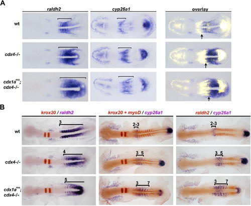
cdx Genes Regulate the Expression Boundaries of raldh2 and cyp26a1. (A) Expression of raldh2 and cyp26a1 in wild-type, cdx4?/? mutants, and cdx-deficient embryos by whole-mount in situ hybridization. Brackets indicate raldh2 and cyp26a1 expression domains in the anterior paraxial mesoderm of 5 somite stage embryos. Right column is an overlay of the raldh2 and cyp26a1 expression patterns, arrows indicate the anterior boundary of the presumptive RA source. (B) Double whole-mount in situ hybridization showing co-staining of krox20, myoD, raldh2, and cyp26a1 at 10 somites in wild-type, cdx4?/? mutants, and cdx-deficient embryos (gene names are color-coded to match their staining product). Brackets and numbers indicate raldh2 and cyp26a1 expression domains and somite position, respectively. EXPRESSION / LABELING:
|
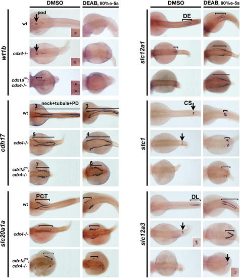
Blocking RA Production in cdx Mutants Partially Rescues Pronephros Position and Distal Segment Formation. (A?C) Expression of cdh17, slc20a1a, and slc12a1 at 48 hpf in wild-type, cdx4?/? mutants, and cdx-deficient embryos that were treated with DMSO (control) or DEAB from 90% epiboly to the 5 somite stage. Lines and numbers indicate expression domains and somite position, respectively. Arrows indicate podocyte and CS positions. Dorsal views are shown, with anterior to the left. Insets show enlarged dorsal views of podocyte (asterisk marks cystic glomeruli which stain weakly) and DL segment staining. |
Zebrafish Proximal Pronephros Segment Domains at 24 hpf. Double in situ hybridizations for segment-restricted genes (purple) and mhc (red). EXPRESSION / LABELING:
|

Zebrafish Distal Pronephros Segment Domains at 24 hpf. Double in situ hybridizations for segment-restricted genes (purple) and mhc (red). EXPRESSION / LABELING:
|
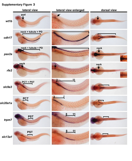
Zebrafish Proximal Pronephros Segment Domains at 48 hpf. Double in situ hybridizations for segment-restricted genes (purple) and mhc (red). EXPRESSION / LABELING:
|

Zebrafish Distal Pronephros Domains at 48 hpf. Double in situ hybridizations for segment-restricted genes (purple) and mhc (red). EXPRESSION / LABELING:
|
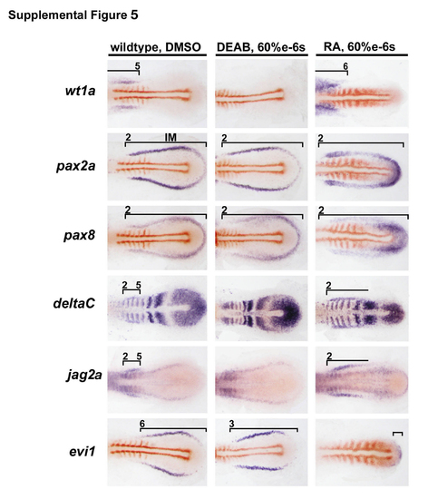
Changes in RA Signaling Cause Early Alterations in Gene Expression Throughout the Intermediate Mesoderm. Expression of intermediate mesodermal markers (purple) and myoD (red) at the 6 somite stage in wild-type embryos incubated with DMSO, DEAB, or 1 x 10⁻⁶ M RA from 60% epiboly until the stages shown, by double in situ hybridization. Brackets indicate expression domains, and numbers indicate somite identity. EXPRESSION / LABELING:
|

Unillustrated author statements |