- Title
-
Zebrafish nephrogenesis involves dynamic spatiotemporal expression changes in renal progenitors and essential signals from retinoic acid and irx3b
- Authors
- Wingert, R.A., and Davidson, A.J.
- Source
- Full text @ Dev. Dyn.
|
Pronephros progenitors are delineated into a series of molecularly distinct regions during early somitogenesis that are retinoic acid (RA) dependent. A–C: Gene expression patterns in the nephron territory in wild-type embryos and lib mutants at the 8 somite stage (A) 15 somite stage (B), and schematized respectively (C,D). Embryos were flat-mounted to remove the yolk and are shown in dorsal views with anterior to the left. Whole-mount in situ hybridization was used to mark kidney gene expression (purple) and the somites with myoD (red). Black lines indicate areas of kidney gene expression and numbers correspond to the somite position. A: In 8 somite wild-types, pax2a transcripts marked all nephron progenitors while dlc and jag2 expression was restricted proximally and mecom expression was restricted distally; a short stretch of overlap between the jag2 and mecom domains was evident. lib embryos had reduced dlc and jag2 domains, and an expanded mecom domain. B: In 15 somite wild-type embryos, hnf1ba marked all nephron progenitors, and the other transcription factors were restricted to subsets of cells in the rostral (hnf4a, hnf1bb), central (irx3b), or caudal (pou3f3b, mecom, emx1, gata3) areas of the nephron territory. lib had a shortened rostral hnf4a area while the central and caudal were expanded. C,D: Schematic depictions of nephron progenitor domains: (top) labeled boxes indicate somite number, and (below) summary of rostral (purple), central (blue) and caudal (green) areas of the nephron progenitors. At 8 somites, the overlap between rostral and caudal areas is depicted as dark purple-colored subset of the rostral area. At 15 somites, subsets of the caudal area are depicted as dark green and light green; gray indicates divergence of nontubule populations (future podocytes and neck segment cells) at the most anterior regions of the renal progenitors. Asterisk marks the posterior limit of the rostral area, which shifted anteriorly in lib at the 8 and 15 somite stages. EXPRESSION / LABELING:
PHENOTYPE:
|
|
Dynamic spatiotemporal expression changes during late somitogenesis precede the appearance of segment identities in the pronephros. Gene expression patterns of transcription factors in nephron progenitors in wild-type embryos at the 20, 22, 24, and 28 somite stage. Embryos are shown in lateral views with anterior to the left, and have been stained by whole-mount in situ hybridization to mark kidney gene expression (purple) and the somites with mhc (red). Black lines indicate areas of high gene expression, and numbers correspond to the somite position. The spatial distributions of hnf1ba, hnf4a, hnf1bb, emx1, and gata3 transcripts were unaltered between 20 and 28 somites of development, although note that the proximal boundary of the emx1 and gata3 domains at 20 somites changed since the 15 somite stage (compare with Fig. 1B). The expression domains of pou3f3a and pou3f3b were altered at 22 and 28 somites, the irx3b and grhl2a expression domains changed at 24 somites, and the etv5 domain was restricted at the 28 somite stage. The expression domain of mecom exhibited multiple alterations between 20 and 28 somites. EXPRESSION / LABELING:
|
|
lib is the most severe genetic model of aldh1a2-deficiency in the zebrafish. A: Lateral views of living 24 and 48 hours postfertilization (hpf) wild-type embryos, nls homozygotes, nls/lib compound heterozygotes, lib homozygotes, and diaminobenzaldehyde (DEAB) -treated wild-types. aldh1a2 mutations are associated with a truncation of the cervical region evident at 24 hpf (arrows indicate the caudal boundary of the otic vesicle [OV] and the rostral boundary of the first somite [S1]). DEAB-treated embryos lack this region, with the OV and S1 located adjacent to each other (indicated by single arrow). At 48 hpf, aldh1a2 mutations are associated with a kink at the head-trunk boundary (black arrow) and pericardial edema (*). Also at 48 hpf, lib and DEAB-treated embryos show pronounced body curvature and develop a lightbulb-shaped yolk compared with wild-types and other aldh1a2 mutants. B: Meiotic mapping placed lib in a genetic interval on linkage group (LG) 7 between z8693 and z11894 that includes aldh1a2. C: Reverse transcriptase-polymerase chain reaction and sequence analysis of aldh1a2 transcripts from lib mutants detected a C → A transversion at nucleotide 174 that is predicted to introduce a Stop codon in lieu of a Tyrosine residue at amino acid position 58 in the 519 amino acid raldh2 enzyme. D: Whole-mount in situ hybridization analysis of aldh1a2 expression (purple) and krox20 expression (red) shows decreased aldh1a2 transcripts both in lib mutants and lib heterozygote embryos, consistent with nonsense mediated decay. EXPRESSION / LABELING:
|
|
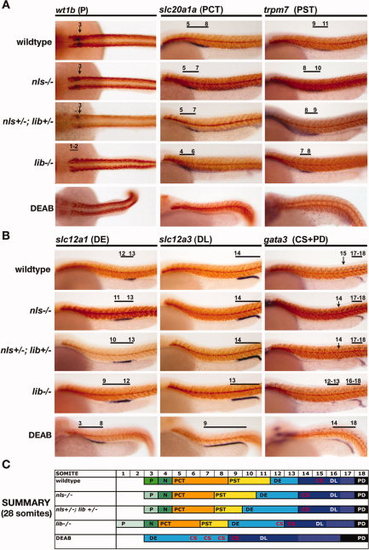
Genetic and chemical models of retinoic acid (RA) biosynthesis deficiency exhibit a similar nephron segment phenotype of reduced proximal fates and expanded distal fates, and lib is the most severely affected aldh1a2 mutant. Whole-mount in situ hybridization analysis for nephron segment markers (purple) and mhc (red) at the 28 somite stage in aldh1a2 mutants and diaminobenzaldehyde (DEAB) -treated wild-type embryos. Embryos are shown in lateral views with anterior to the left, with the exception of dorsal views to show podocytes. Black brackets and arrows indicated expression domains, and numbers correspond to somite position. A,B: Proximal fates of podocytes, the proximal convoluted tubule (PCT) and proximal straight tubule (PST), were all reduced in the absence of RA biosynthesis (A), whereas distal fates of the distal early (DE), distal late (DL), corpuscle of Stannius (CS), and pronephric duct (PD) were expanded (B). C: Summary of nephron segmentation with respect to embryo somite number. Each color represents a different epithelial population: podocytes (green), PCT (orange), PST (yellow), DE (light blue), CS (red lettering indicates location among nephron tubule cells), DL (dark blue), and PD (black), and the overlap between DL and PD-expressed genes is indicated in white. The nephron segment phenotypes among RA-deficient zebrafish embryos showed a graded severity such that: nls homozygote < nls/lib compound heterozygotes < lib homozygotes < DEAB-treated wild-type embryos. EXPRESSION / LABELING:
PHENOTYPE:
|
|
tbx16-dependent paraxial mesoderm formation is essential for proximodistal nephron patterning. Whole-mount in situ hybridization analysis for nephron segment markers (purple) and mhc (red) at the 28 somite stage in wild-type embryos and tbx16 morpholino (MO) -injected embryos. Embryos are shown in lateral views with anterior to the left. Black brackets and arrows indicate expression domains, and numbers correspond to somite position. A: tbx16 knockdown in wild-types was associated with formation of reduced podocyte numbers, shown by reduced wt1b expression, and shorter proximal segments, shown by smaller nbc1, slc10a1a, and trpm7 gene expression domains; conversely, distal segments were expanded, shown by longer clck, slc12a1, and slc12a3 expression domains. The DL (gata3-expressing segment) was unaffected. B: tbx16 morpholino injected animals have a reduced rostral domain (hnf4a), and shifts in the central (irx3b), and caudal (mecom) domains at 28 somites that are consistent with shorter proximal and expanded distal segments. C,D: tbx16 morpholino injected animals have dramatically reduced expression of aldh1a2 at 28 somites (C) and 5 somites (D). EXPRESSION / LABELING:
PHENOTYPE:
|
|
irx3b activity is requisite for distal early (DE) segment identity, and the absence of retinoic acid (RA) and irx3b activity generates nephrons with a distal late (DL) -only tubule identity. A: Whole-mount in situ hybridization analysis for nephron segment markers (purple) and mhc (red) at the 28 somite stage in wild-type embryos, irx3b morpholino (MO) -injected embryos, lib homozygous mutants, lib homozygous mutants injected with irx3b MO, diaminobenzaldehyde (DEAB) -treated embryos, and DEAB-treated embryos injected with irx3b MO. Embryos are shown in lateral views with anterior to the left. Black brackets and arrows indicated expression domains, and numbers correspond to somite position. irx3b knockdown in wild-types and lib mutants was associated with DE segment abrogation, expansion of the proximal convoluted tubule (PCT) and proximal straight tubule (PST) segments as well as the corpuscle of Stannius (CS) population. In DEAB-treated embryos, which normally only form DE and DL tubule segments, irx3b knockdown was associated with expansion of the DL while the PD is unchanged. B: In DEAB-treated embryos, irx3b expression marked the DE domain that becomes defined by expression of slc12a1. DEAB-treated embryos injected with irx3b MO showed expression of irx3b in the same region of the pronephros, suggesting that DE regionalization had been established, but that in the absence of irx3b DE-specific solute transporter expression does not ensue. C: Summary of nephron segmentation with respect to embryo somite number. Asterisks in the proximal DL of DEAB-treated wild-types injected with irx3b MO indicate partial regionalization that is blocked/prevented in these animals. EXPRESSION / LABELING:
PHENOTYPE:
|
|
Cell proliferation of pronephros progenitors is not required to establish segment boundaries. Whole-mount in situ hybridization analysis for nephron segment markers (purple) and mhc (red) at the 28 somite stage in wild-type embryos treated with dimethyl sulfoxide (DMSO; vehicle alone; A) or camptothecin starting at the 24, 22, 20, 18, and 15 somite stage or nocodazole starting at the 22 somite stage (B). Embryos are shown in lateral views with anterior to the left. Black brackets and arrows indicated expression domains, and numbers correspond to somite position. Embryos in all chemical treatments formed proximal convoluted tubule (PCT), proximal straight tubule (PST), distal early (DE), and distal late (DL) segments at the same somite position as wild-type embryos, consistent with a negligible role for cell proliferation in the establishment of segment domains after the 15 somite stage. The segments detected using the following transcripts for all embryos except the 15 somite stage treatment: PCT by slc20a1a, PST by trpm7, DE by slc12a1, DE by slc12a3. The 15 somite stage camptothecin-treated embryos were significantly delayed by the drug treatment and did not show trpm7 or slc12a1 transcripts (data not shown). To probe the existence of these areas, the overlap between hnf4a and irx3b was ascertained, as indicated by a single asterisk for hnf4a and double asterisk for irx3b transcripts. The overlap between hnf4a and irx3b showed the presence of the PCT identity (cells that expressed hnf4a only) and PST (region of cells that expressed both hnf4a and irx3b), a correlation based on that seen in wild-type embryos at this stage. |
|
lib mutant morphology and nephron segmentation are rescued by treatment with exogenous retinoic acid (RA) or overexpression of aldh1a2. A: Lateral views of living 28 somite wild-type and lib embryos compared with embryos injected with aldh1a2 cRNA transcripts. lib embryos formed a normal body trunk without curvature and a kink between the head and trunk, although mild pericardial edema still developed. B: Table of rescue percentages in offspring from nls, nls/lib, and lib heterozygous crosses after treatment with dimethyl sulfoxide (DMSO; vehicle alone), all-trans RA at 1 × 10⁻⁹M, all-trans RA at 1 × 10⁻⁸M, and wild-type aldh1a2 cRNA injection. Rescue evaluation was determined by presence of pectoral fins in mutant embryos. lib homozygote embryos were rescued only by higher RA or aldh1a2 overexpression, while nls homozygotes and nls/lib compound heterozygotes were rescued at the lower RA dosage. C: Whole-mount in situ hybridization analysis for nephron segment markers (purple) and mhc (red) at the 28 somite stage in wild-type and lib mutants after exogenous treatment with all-trans RA at 1 × 10⁻⁹M. Embryos are shown in lateral views with anterior to the left, with the exception of dorsal views to show podocytes. Black brackets and arrows indicated expression domains, and numbers correspond to somite position. RA treatment restored the podocyte population in lib, and expanded proximal tubule fates (proximal convoluted tubule [PCT] and proximal straight tubule [PST]), while the distal fates (distal early [DE], distal late [DL], pronephric duct [PD]) were all reduced, showing pronephros progenitors were competent to respond to elevated exogenous RA. Similar phenotype trends were observed in wild-types exposed to this dosage of RA. D: Summary of nephron segmentation with respect to embryo somite number in wild-type and lib with DMSO or all-trans RA 1 × 10⁻⁹M exposure. |
|
Alterations in retinoic acid receptor-alpha (RAR-α) activity are necessary and sufficient to alter proximodistal nephron patterning. A,B: Whole-mount in situ hybridization analysis for nephron segment markers (purple) and mhc (red) at the 28 somite stage in wild-type embryos treated with dimethyl sulfoxide (DMSO; vehicle only), an RAR-α antagonist, or RAR-α agonist. Embryos are shown in lateral views with anterior to the left, with the exception of dorsal views to show podocytes. Black brackets and arrows indicated expression domains, and numbers correspond to somite position. RAR-α antagonist treatment reduced proximal segments and expanded distal segments, while RAR-α agonist treatment expanded proximal segments at the expense of distal fates. C: Summary of nephron segmentation with respect to embryo somite number for each treatment. |
|
Dynamic alterations in renal transcription factors in lib mutants follow trends similar to wild-type embryos in rostral and central domains. Gene expression patterns of transcription factors in nephron progenitors in wild-type embryos compared with lib mutant embryos at the 20, 22, 24, and 28 somite stage. Embryos are shown in lateral views with anterior to the left, and have been stained by whole-mount in situ hybridization to mark kidney expression (purple) and the somites with mhc (red). Black lines indicate areas of high gene expression, and numbers correspond to the adjacent somite position. The spatial distributions of hnf1a, hnf4a, and hnf1bb transcripts were unaltered between 20 and 28 somites (28 somite stage not shown). The distributions of etv5 and irx3b changed in wild-types and lib at the 28 and 24 somite stages, respectively. |
|
Dynamic alterations in renal transcription factors in lib mutants show some trends analogous to wild-type embryo caudal domains, but with some differences. Gene expression patterns of transcription factors in nephron progenitors in wild-type embryos compared with lib mutant embryos at the 20, 22, 24, and 28 somite stage. Embryos are shown in lateral views with anterior to the left, and have been stained by whole-mount in situ hybridization to mark kidney expression (purple) and the somites with mhc (red). Black lines indicate areas of high gene expression, and numbers correspond to the adjacent somite position. The spatial distributions of pou3f3a and pou3f3b transcripts in lib mutants were continually expressed in rostral regions between 20 and 28 somites (28 somite stage not shown). The distribution of mecom changed progressively in both wild-types and lib throughout the 20–28 somite stages. The distribution of emx1 and gata3 transcripts was unaltered in wild-types between the 20 and 28 somite stages, whereas lib had progressive restrictions of these transcripts to caudal populations. |
|
Transcription factor domain alterations in irx3b morphants correspond to changes in segment boundaries. A: Gene expression patterns of transcription factors in nephron progenitors in wild-type embryos and irx3b morpholino (MO) -injected animals at the 28 somite stage. Embryos are shown in lateral views with anterior to the left, and have been stained by whole-mount in situ hybridization to mark kidney gene expression (purple) and the somites with mhc (red). Black lines indicate areas of high gene expression, and numbers correspond to the somite position. In irx3b morphants, the domains of hnf1ba, hnf1g, irx3b, mecom, emx1, and gata3 were unchanged, while the expression of hnf4a was expanded slightly and the rostral boundary of pou3f3a and pou3f3b were shifted distally by one somite. In addition, the pou3f3a domain was expanded to somite 14. B: The domain of transcription factor gene expression in wild-type and irx3b morphant embryos correlates closely to the segment pattern. (Foreground) Summary of transcription factor expression domains (represented by black bars) are schematized in the nephrons at the 28 somite stages (Note: wild-type schema is repeated from Fig. 3 for easy comparison). (Background) Nephron segment identities form adjacent to particular somites (demarcated by numbered columns, top), and each segment region is color-coded to envisage a comparison between the transcription factor expression domains and the nephron segments. Segment color codes are N, green; PCT, orange; PST, yellow; DE, light blue; DL, dark blue; and PD, gray. |


