
Fig. S1
- ID
- ZDB-IMAGE-110811-26
- Publication
- Wingert et al., 2011 - Zebrafish nephrogenesis involves dynamic spatiotemporal expression changes in renal progenitors and essential signals from retinoic acid and irx3b
- All Figures
- Figures for Wingert et al., 2011
|
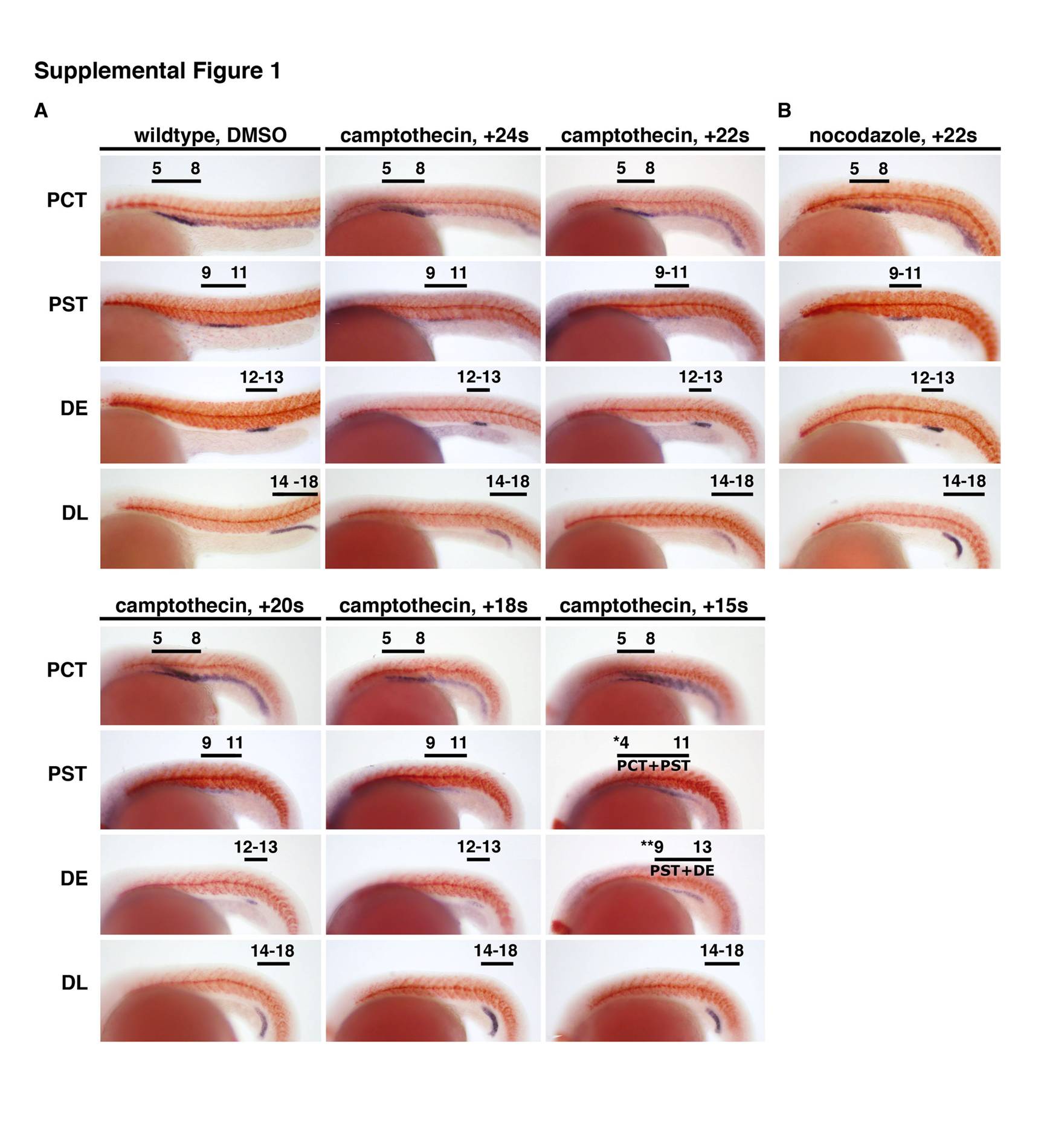
Fig. S1 Cell proliferation of pronephros progenitors is not required to establish segment boundaries. Whole-mount in situ hybridization analysis for nephron segment markers (purple) and mhc (red) at the 28 somite stage in wild-type embryos treated with dimethyl sulfoxide (DMSO; vehicle alone; A) or camptothecin starting at the 24, 22, 20, 18, and 15 somite stage or nocodazole starting at the 22 somite stage (B). Embryos are shown in lateral views with anterior to the left. Black brackets and arrows indicated expression domains, and numbers correspond to somite position. Embryos in all chemical treatments formed proximal convoluted tubule (PCT), proximal straight tubule (PST), distal early (DE), and distal late (DL) segments at the same somite position as wild-type embryos, consistent with a negligible role for cell proliferation in the establishment of segment domains after the 15 somite stage. The segments detected using the following transcripts for all embryos except the 15 somite stage treatment: PCT by slc20a1a, PST by trpm7, DE by slc12a1, DE by slc12a3. The 15 somite stage camptothecin-treated embryos were significantly delayed by the drug treatment and did not show trpm7 or slc12a1 transcripts (data not shown). To probe the existence of these areas, the overlap between hnf4a and irx3b was ascertained, as indicated by a single asterisk for hnf4a and double asterisk for irx3b transcripts. The overlap between hnf4a and irx3b showed the presence of the PCT identity (cells that expressed hnf4a only) and PST (region of cells that expressed both hnf4a and irx3b), a correlation based on that seen in wild-type embryos at this stage.