
Fig. 4
- ID
- ZDB-IMAGE-110811-22
- Genes
- Publication
- Wingert et al., 2011 - Zebrafish nephrogenesis involves dynamic spatiotemporal expression changes in renal progenitors and essential signals from retinoic acid and irx3b
- All Figures
- Figures for Wingert et al., 2011
|
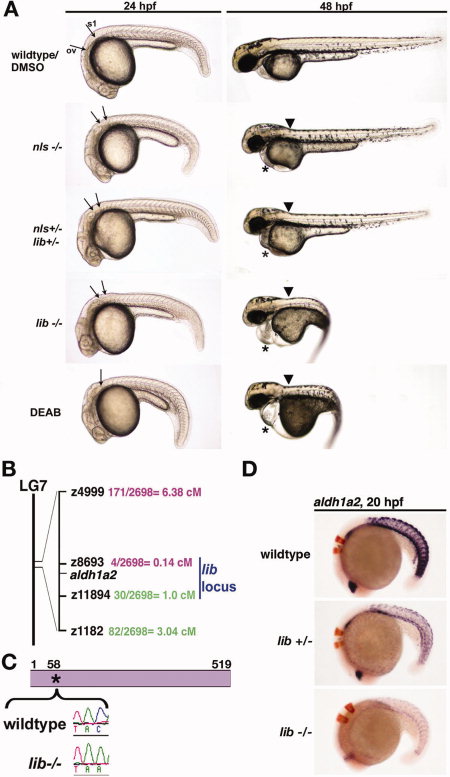
Fig. 4 lib is the most severe genetic model of aldh1a2-deficiency in the zebrafish. A: Lateral views of living 24 and 48 hours postfertilization (hpf) wild-type embryos, nls homozygotes, nls/lib compound heterozygotes, lib homozygotes, and diaminobenzaldehyde (DEAB) -treated wild-types. aldh1a2 mutations are associated with a truncation of the cervical region evident at 24 hpf (arrows indicate the caudal boundary of the otic vesicle [OV] and the rostral boundary of the first somite [S1]). DEAB-treated embryos lack this region, with the OV and S1 located adjacent to each other (indicated by single arrow). At 48 hpf, aldh1a2 mutations are associated with a kink at the head-trunk boundary (black arrow) and pericardial edema (*). Also at 48 hpf, lib and DEAB-treated embryos show pronounced body curvature and develop a lightbulb-shaped yolk compared with wild-types and other aldh1a2 mutants. B: Meiotic mapping placed lib in a genetic interval on linkage group (LG) 7 between z8693 and z11894 that includes aldh1a2. C: Reverse transcriptase-polymerase chain reaction and sequence analysis of aldh1a2 transcripts from lib mutants detected a C → A transversion at nucleotide 174 that is predicted to introduce a Stop codon in lieu of a Tyrosine residue at amino acid position 58 in the 519 amino acid raldh2 enzyme. D: Whole-mount in situ hybridization analysis of aldh1a2 expression (purple) and krox20 expression (red) shows decreased aldh1a2 transcripts both in lib mutants and lib heterozygote embryos, consistent with nonsense mediated decay.