
- Title
-
Knockout of longevity gene Sirt1 in zebrafish leads to oxidative injury, chronic inflammation, and reduced life span
- Authors
- Kim, D.H., Jung, I.H., Kim, D.H., Park, S.W.
- Source
- Full text @ PLoS One
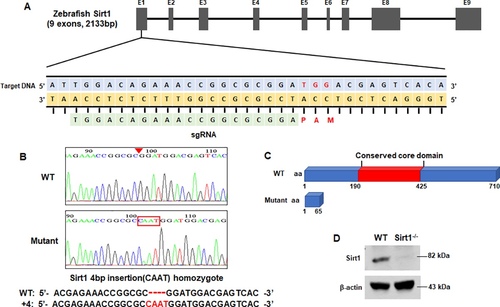
Sirt1 Knockout by Crispr/Cas9 technique. (A) Schematic illustration of the Sirt1 locus. Exon 1 of the Sirt1 gene was targeted. The CRISPR target site and PAM motif are indicated. (B) Sequence confirmation of four-nucleotide insertion mutations. (C) Predicted truncation of the SIRT1 protein. (D) Expression of the Sirt1 mutant was verified by western blot using an antibody of Sirt1; β-actin was used as a loading control. |

Short term phenotype of Sirt1 -/- mutants. Four-days post fertilization embryos were used to obtain representative images of short term phenotypes of wildtype and Sirt1-/- mutants. (A) Phenotypes of wildtype and Sirt1-/- zebrafish embryos. Note there are no differences in appearance between wildtype and Sirt1-/- mutant embryos. (B) The embryo survival curves of wildtype and Sirt1-/- mutants after treatment with 0.5 or 1 mM tBOOH. Note that wildtype embryos died earlier than Sirt1-/- mutants. (C) ROS levels in 0.5 mM tBOOH-treated wildtype and Sirt1-/- embryos were examined by H2DCFDA fluorescence. (D) Detection of apoptosis in 0.5 mM tBOOH-treated wildtype and Sirt1-/- mutant embryos by TdT-mediated dUTP nick end labeling (TUNEL). Note that Sirt1-/- mutants showed higher levels of ROS and TUNEL than wildtype embryos. The effect of NAC treatment on ROS content and apoptosis incidence was studied by H2DCFDA fluorescence (E) and the TUNEL assay (F). Embryos were exposed to treatment with NAC treatment for 24 h. Results showed that TUNEL-positive cells increased in both, tBOOH-treated wildtype and Sirt1-/-groups; furthermore, NAC treatment was effective for both groups. |
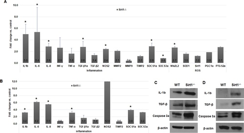
Quantitative RT-PCR and western blot analysis. Samples were prepared from (A, C) 4-dpf whole embryos, and (B, D) internal organs from 3-month-old zebrafish. Data are means, and bars indicate standard error. (A, B) Quantitative RT-PCR showed higher upregulation of inflammation-related genes in Sirt1-/- than in controls. (C, D) Western blot hybridization showed upregulation of IL-1b, active-caspase 3 and TGF-β. Control, wildtype; IL-1b, 18kDa; caspase 3a, 29 and 31 kDa; TGF-β, 45 kDa; β–actin, 43 kDa. *P < 0.05 versus control. |

Microscopic observations of wildtype and Sirt1-/- adult zebrafish. H&E images of wildtype and Sirt1-/- zebrafish aged 6, 12, and 18 months. Sirt1-deficiency caused intestinal atrophy and inflammatory cell infiltration PHENOTYPE:
|
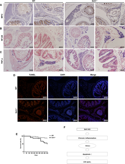
Expression of genes involved in inflammation and apoptosis induced by Sirt1-knockout induced inflammation. All images were obtained from 12-month-old zebrafish. (A) Immunohistochemical staining for myeloperoxidase (MPO). (B) ISH for NF-kB and (C) TNF-α expression. (D) TUNEL assay with 18-month old zebrafish organ sections. Wildtype organ section with few apoptotic cells and Sirt1-/- organ section with numerous apoptotic cells, as per the TUNEL assay. (E) Survival curves of wildtype and Sirt1-/- mutant zebrafish. Kaplan–Meier analysis showed poor survival of Sirt1-/- compared to the wildtype. (n = 100 zebrafish in each group). Survival curves lasted for 540 days (observation started when zebrafish were 3 months old.) and were statistically compared using the Log-Rank Test. (F) Schematic diagram of the major findings of this study. Sirt1-/—induced chronic inflammation led to the activation of oxidative stress and apoptosis. The continuous repetition of these events reduced the lifespan of zebrafish. |