|
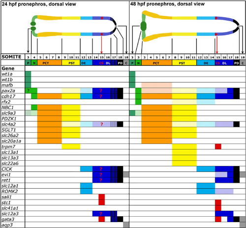
Pronephros Expression of Known Renal Genes and Solute Transporters Isolated Using Functional Genomic. The domain of each pronephros-expressed gene was mapped along the embryonic axis according to somite boundaries, using double in situ hybridization with mhc to label the myotomes. Solid color bars indicate strong expression, light color bars indicate weak expression, and blue/black stripe bars indicate overlap in the expression domains of DL and PD markers; note that rfx2 expression at 48 hpf is discontinuous in the PST and DE domains. Schematic anatomy of the pronephros at 24 hpf (top, left) compared to 48 hpf (top, right). The cloaca (C) is not considered a segment of the pronephros, but is shown to depict the terminus of the pronephros.
|