|
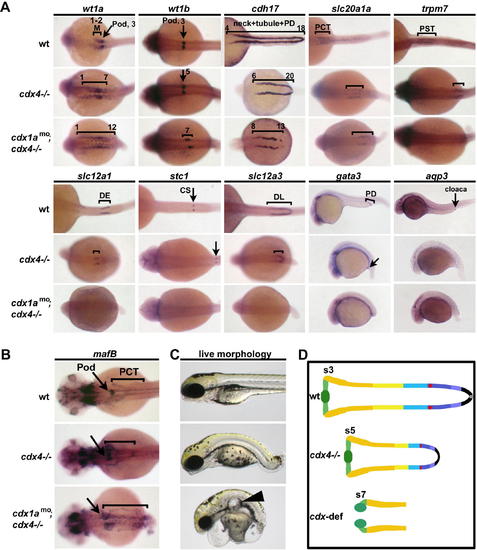
cdx Genes Position the Pronephros along the Embryonic Axis and Are Requisite for Distal Segment Formation. Expression of segment markers was performed on wild-type, cdx4–/– mutants, and cdx-deficient embryos at (A) 26 hpf and (B) 48 hpf using whole-mount in situ hybridization. Brackets and arrows indicate expression domains in each embryo. Numbers indicate somite number. Dorsal views of embryos, with anterior to the left, with the exception of lateral views for gata3 and aqp3 expression. (C) Live wild-type, cdx4–/– mutants, and cdx-deficient embryos at 72 hpf. Arrowhead indicates glomerular cyst in the cdx-deficient embryo. Lateral views are shown, with anterior to the left. (D) Summary of nephron segmentation in cdx4–/– mutants and cdx-deficient embryos as compared to wild-type at 48 hpf. Numbers indicate anterior somite boundary of each pronephros.
|