
Fig. 7
- ID
- ZDB-FIG-071116-6
- Publication
- Wingert et al., 2007 - The cdx Genes and Retinoic Acid Control the Positioning and Segmentation of the Zebrafish Pronephros
- Other Figures
- All Figure Page
- Back to All Figure Page
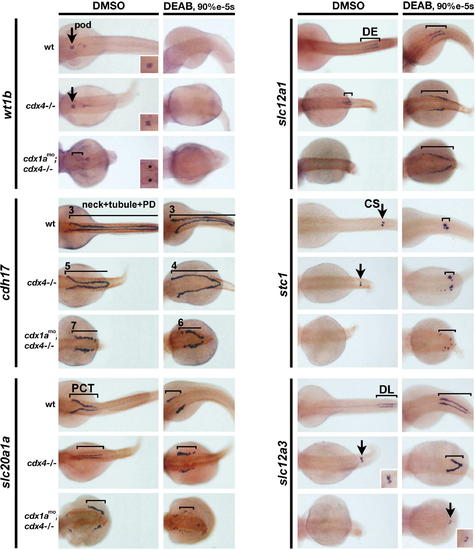
Blocking RA Production in cdx Mutants Partially Rescues Pronephros Position and Distal Segment Formation. (A–C) Expression of cdh17, slc20a1a, and slc12a1 at 48 hpf in wild-type, cdx4–/– mutants, and cdx-deficient embryos that were treated with DMSO (control) or DEAB from 90% epiboly to the 5 somite stage. Lines and numbers indicate expression domains and somite position, respectively. Arrows indicate podocyte and CS positions. Dorsal views are shown, with anterior to the left. Insets show enlarged dorsal views of podocyte (asterisk marks cystic glomeruli which stain weakly) and DL segment staining. |
| Genes: | |
|---|---|
| Fish: | |
| Condition: | |
| Knockdown Reagent: | |
| Anatomical Terms: | |
| Stage: | Long-pec |
| Fish: | |
|---|---|
| Condition: | |
| Knockdown Reagent: | |
| Observed In: | |
| Stage: | Long-pec |