- Title
-
The small molecule Mek1/2 inhibitor U0126 disrupts the chordamesoderm to notochord transition in zebrafish
- Authors
- Hawkins, T.A., Cavodeassi, F., Erdelyi, F., Szabo, G., and Lele, Z.
- Source
- Full text @ BMC Dev. Biol.
|
U0126 application during a specific time-window decreases ERK phosphorylation specifically. A: Diphospho-Erk staining in the trunk of wt 24 hpf embryos.B: U0126 eliminates ERK1/2 phosphorylation (green) in the trunk and tail of 24 hpf zebrafish embryos. Notice that U0126-treated embryos have been overexposed (can be judged by the autofluorescence of the yolk sac extension) and still fail to show specific staining. C: U0124 does not eliminate the p-Erk staining. D: PD98059 also does not eliminate p-Erk staining when given just below the level of lethal toxicity (25 μM). E: Somewhat weaker but still significant p-Erk staining in embryos treated with U0126 starting at 10 hpf. F: Western-blot analysis demonstrated a slight but incomplete reduction in pErk1/2 levels in 10-somite embryos.G: Stage and dose-dependency of U0126 application. There is a dramatic drop in the percentage of affected embryos when treatment is applied after epiboly is completed. H: Washout experiments reveal the necessity of U0126 to be present until at least the 18 somite stage to produce the full phenotype. |
|
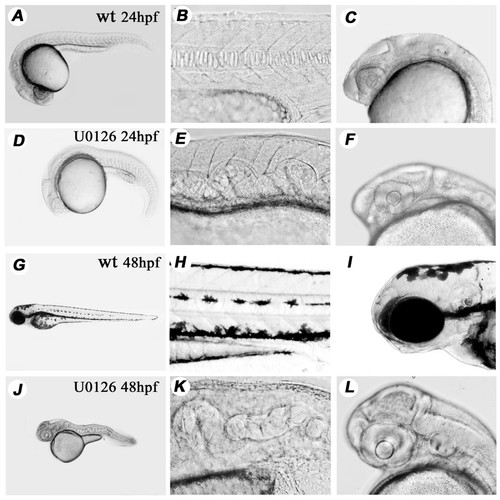
U0126 produces a specific and consistent phenotype affecting pigmentation and notochord development. Phenotype of wild type and U0126-treated embryos at 24 hpf. (A-F) and at 48 hpf (G-L). There is a total lack of pigmentation including in the RPE in the eye (compare panels I and L), twists and undulations in the NC are already evident at 24 hpf. (D, E) and the trunk and tail of treated embryos is shortened at 48 hpf (compare panels G and J). The large yolk sac in 48 hpf embryos indicates that circulation is not established properly to carry away its content (see also later in Fig. 5) Panels A-C, G-I: wild type embryos. Panels D-F, J-L: U0126-treated embryos. |
|
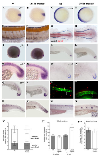
Early processes are not affected by U0126 with the exception of apoptosis. A-D: Expression the dorsal marker gsc (A, B,), and the ventral mesodermal marker gene eve1 (D, E) is not changed in 7 hpf. U0126-treated animals. As treated and wt animals could not be separated at this stage, a large number of embryos were treated and some of them were raised till 24 hpf. to make sure the drug was effective. E, F: Narrower, straighter somites in U0126-treated embryos as demonstrated by the reduced distance between the motoneurons (1 motoneuron/somite) stained with α-Acetylated-Tubulin antibody. G, H: Lateral mesoderm marker gene pax 2.1 (purple) and the somitic mesodermal marker gene myoD (red) do not show altered expression in dorsal view of flat-mounted 9-somite stage embryos. Expression of genes involved in tail development is not changed after U0126-treatment. I-L:flh. M, N: cdx1. O.P: eve1. Q, R: fgf8. S, T: Examples of shh-GFP and α-phosphorylated Histone3 double antibody staining used to count mitotic cells in the NC. U-X: Examples of TUNEL stainings of 24 hpf (U, V) and 48 hpf (W, X) wt and U0126-treated embryos (inset in panel X shows larger magnification and see these cells also in Fig. 6/F marked with yellow arrows). Y′: The average numbers of phosphorylated Histone3 (pH3) positive cells in the anterior and posterior trunk and tail do not differ between wt and U0126-treated embryos. Average numbers of TUNEL positive cells in whole embryos (Y″) and notochord-only (Y′″) at 20 somites and 24 hpf demonstrate a significant increase in U0126-treated embryos at 24 hpf (p = 0.0087 unpaired t-test with welch's correction, 24 hpf values). Note that there is no change in apoptosis within the NC. Panels A, C, E, G, I, K, M, O, Q, S, U, W: wild type embryos. Panels B, D, F, H, J, L, N, P, R, T, V, X: U0126-treated embryos. Panels A, D animal pole view, dorsal to the right. |
|
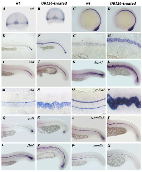
Genes important in notochord differentiation exhibit two types of regulatory pattern during chordamesoderm to mature NC transition. A-H: Expression of ntl is normal during early NC development (A-D) in treated embryos but fails to get downregulated during the chordamesoderm to mature NC transition step (E-H). (A, B: 7 hpf; C, D: 12 hpf; E-H 24 hpf.) This pattern of developmental arrest occurs in ehh (I, J), shh (L, K), col2α1 (M, N) and hsp47 (O, P) expression. Q-X: mRNA levels of fkd1 (Q, R), fkd4 (S, T), spondin2 (U, V) and mindin (W, X) however decreases normally upon chordamesoderm to mature NC transition. Notice the unchanged expression of marker genes in the floor plate (M-X) and in the hypochord (O, P, U-X) of U0126-treated embryos. Panels: A, C, E, G, I, K, M, O, Q, S, U, W: wild type. Panels: B, D, F, H, J, L, N, P, R, T, V, X: U0126-treated embryos. |
|
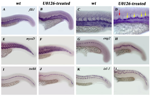
Not all tissues receiving notochord-emitted signals are affected by U0126 treatment. A-D: flk-1 expression reveals defects in dorsal aorta differentiation (green arrow in panel D) and in the sprouting of intersomitic vessels. Notice that both lack of sprouting (yellow arrows) and double sprouting (red arrow) occurs in U0126-treated embryos (D). Expression of the early muscle differentiation marker myoD is normal in treated embryos (E, F) but the muscle pioneer cell marker eng2 is upregulated (G, H). The floor plate of the neural tube differentiates normally (I, J and also Fig. 4/M–X). Isl-1 expression shows that the sensory Rohon-Beard neurons and motoneurons both develop properly in treated embryos (K, L) |
|
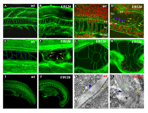
Defects in the cellular organization of the notochord and PNBM structure of U0126-treated embryos. In 24 hpf. U0126-treated embryos notochordal cells adhere to the sheath at preferential focal points which results in the bending of the NC (red arrow in panel B). Phalloidin stainings reveal smaller amount of fibrillar actin present between NC cells but not in the overlying neural tube. Notice the large vacuolar cells and the periferial positioning of nuclei in U0126-treated embryos (panels C, D). By 48 hpf. the NC of U0126-treated embryos with milder phenotypes contains smaller rounded cells which become apoptotic (yellow arrows in panel F and see also in Fig. 3/X). Embryos with stronger phenotypes have large bulges protruding where the flat sheath cells surrounding the large vacuolated cells are missing (see blue arrows in panel E for normal sheath cells and in panel H marking the last sheath cells surrounding the bulge; red arrows mark the region devoid of sheath cells). Laminin expression and ultrastructure of the inner layer of the PNBM (marked with blue arrowheads) composed mainly of laminins are normal in NC treated embryos as demonstrated by whole-mount immunhistochemistry (I, J) and TEM pictures (K, L). The middle (green bracket) and outer (yellow bracket) layers of the PNBM composed mainly of circumferential and longitudinal collagen fibers respectively show decreased length and/or disorganization in the orientation of fibers (K, L). Panels A, C, E, G, I, K: wild type embryos. Panels B, D, F, H, J, L: U0126-treated embryos. |
|
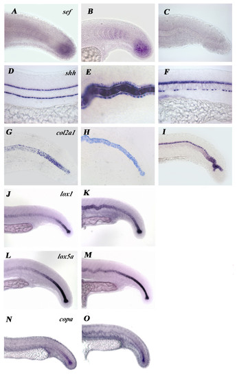
Inhibition of the FGF pathway by SU5402 does not show any similarities to U0126-treated embryos. A-C: Expression of the Fgf-pathway target gene sef is downregulated in SU5402 (C) but not in U0126-treated (B) embryos. D-I: Expression of the notochordal marker genes col2a1 (D-F) and shh (G-I) also show different defects in SU5402 and U0126-treated embryos. J-M: Expression of lox1 (J, K) and lox5b (L-M) is not decreased normally by U0126-treatment. N, O: copa mRNA persists and is not downregulated by U0126-treatment. |


