Fig. 3
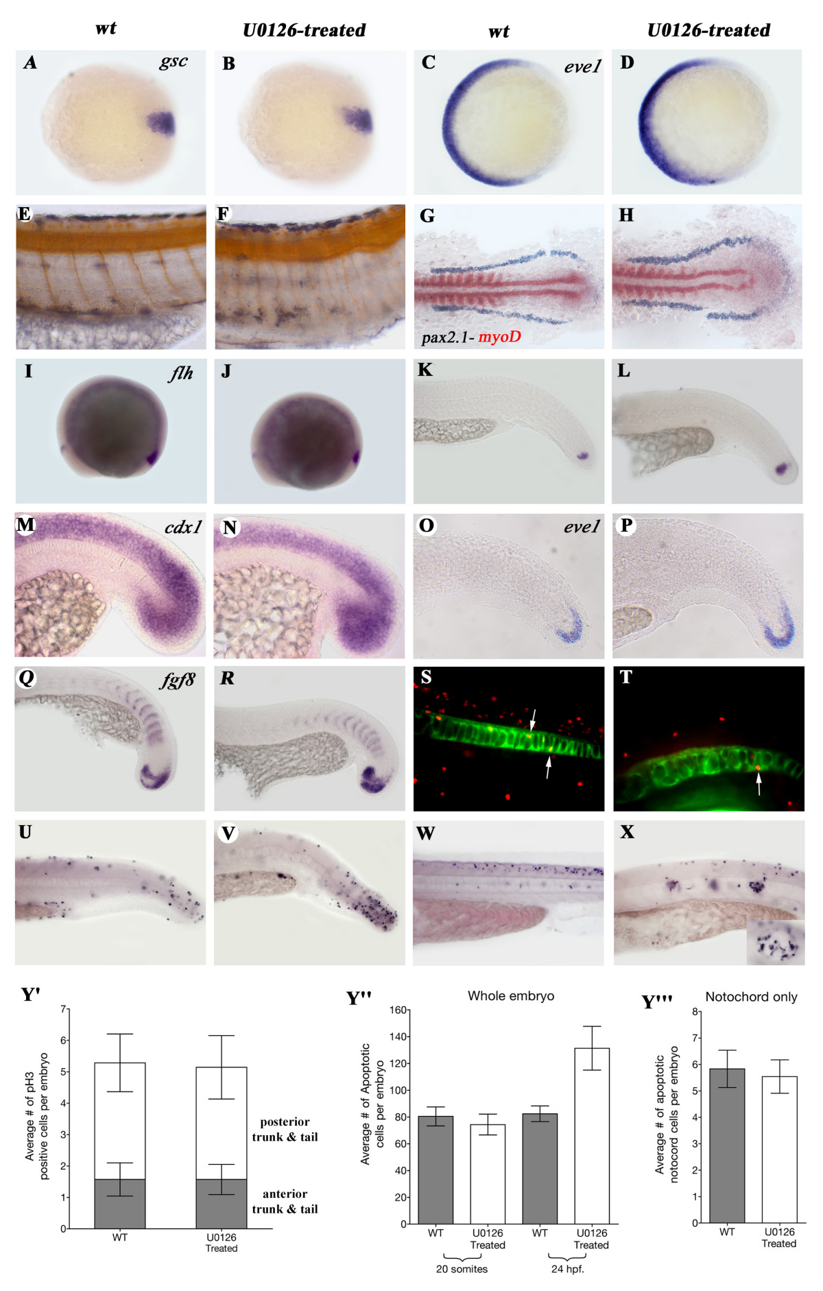
Fig. 3 Early processes are not affected by U0126 with the exception of apoptosis. A-D: Expression the dorsal marker gsc (A, B,), and the ventral mesodermal marker gene eve1 (D, E) is not changed in 7 hpf. U0126-treated animals. As treated and wt animals could not be separated at this stage, a large number of embryos were treated and some of them were raised till 24 hpf. to make sure the drug was effective. E, F: Narrower, straighter somites in U0126-treated embryos as demonstrated by the reduced distance between the motoneurons (1 motoneuron/somite) stained with α-Acetylated-Tubulin antibody. G, H: Lateral mesoderm marker gene pax 2.1 (purple) and the somitic mesodermal marker gene myoD (red) do not show altered expression in dorsal view of flat-mounted 9-somite stage embryos. Expression of genes involved in tail development is not changed after U0126-treatment. I-L:flh. M, N: cdx1. O.P: eve1. Q, R: fgf8. S, T: Examples of shh-GFP and α-phosphorylated Histone3 double antibody staining used to count mitotic cells in the NC. U-X: Examples of TUNEL stainings of 24 hpf (U, V) and 48 hpf (W, X) wt and U0126-treated embryos (inset in panel X shows larger magnification and see these cells also in Fig. 6/F marked with yellow arrows). Y′: The average numbers of phosphorylated Histone3 (pH3) positive cells in the anterior and posterior trunk and tail do not differ between wt and U0126-treated embryos. Average numbers of TUNEL positive cells in whole embryos (Y″) and notochord-only (Y′″) at 20 somites and 24 hpf demonstrate a significant increase in U0126-treated embryos at 24 hpf (p = 0.0087 unpaired t-test with welch's correction, 24 hpf values). Note that there is no change in apoptosis within the NC. Panels A, C, E, G, I, K, M, O, Q, S, U, W: wild type embryos. Panels B, D, F, H, J, L, N, P, R, T, V, X: U0126-treated embryos. Panels A, D animal pole view, dorsal to the right.

