
- Title
-
β-Amyloid precursor protein-b is essential for Mauthner cell development in the zebrafish in a Notch-dependent manner
- Authors
- Banote, R.K., Edling, M., Eliassen, F., Kettunen, P., Zetterberg, H., Abramsson, A.
- Source
- Full text @ Dev. Biol.
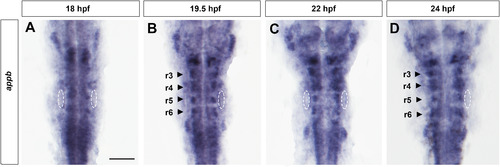
Expression pattern of appb in the zebrafish hindbrain. The appb mRNA transcript pattern during development revealed by whole-mount in situ hybridization. Dorsal views of flat-mounted wild-type embryos, anterior to the top, at the indicated stages (A-D). Arrowheads indicate rhombomere position. r3-r6, rhombomeres 3-6 and dotted oval circles indicate the otocyst. Scale bar, 75 µm. EXPRESSION / LABELING:
|

Knockdown of Appb inhibit Mauthner cell development. Whole-mount immunostaining of embryos at 48 hpf (A-C). Confocal images (anterior to the top) of hindbrain stained with anti-neurofilament RMO44 antibody marking reticulospinal neurons in control MO (A) and appb MO injected (B,B2) embryos. Note the 1-cell/no-cell phenotype in morphants. The large M-cells (M, in A) are located in r4. The absent M-cell is noted by an asterisk (B,B′). M-cell number is rescued in embryos coinjected with appb MO and appb mRNA (C). Cell counts of M-cell number (D). M, Mauthner cell; n, number of embryos and r3-r6, rhombomeres 3-6. Scale bar, 50 µm. |

Appb morphants show defect in eliciting normal behavior. Schematic representation of the behavioral setup with an immobilized 2 dpf embryo in agarose with tail remaining free (A). One electrode was placed superficially over the head region and the other negative electrode into the agarose. Quantification of the tail response in control and appb MO embryos (B). Graph represents data as the mean of three trials for each animal. ****p<0.0001. PHENOTYPE:
|

Decreased neuronal differentiation in Appb morphants. Dorsal view of the flat-mounted control MO (A-C) and appb MO (D-F) injected embryos (anterior at the top) at 24 hpf showing neurog1 (A, D), deltaA (B, E) and deltaD (C, F) expression levels in the hindbrain. Scale bar, 75 µm. |
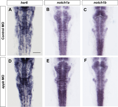
appb knockdown leads to increased Notch signaling. Dorsal view of flat-mounted control MO (A-C) and appb MO (D-F) injected embryos (anterior at the top) at 24 hpf showing her6 (A, D), notch1a (B, E) and notch1b (C, F) expression levels in the hindbrain. Scale bar, 75 µm. |
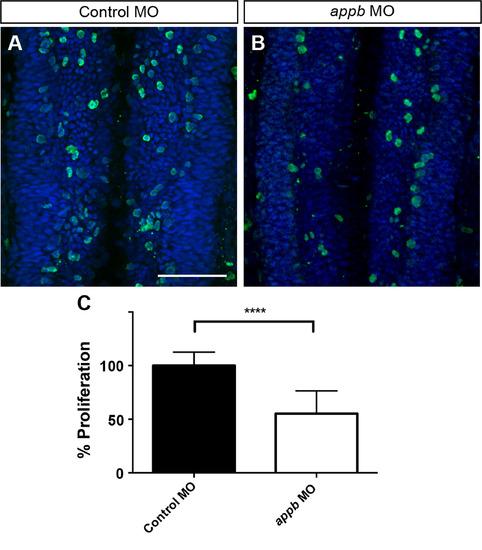
Decreased proliferation following appb knockdown. Whole-mount immunostaining of embryos at 24 hpf (A-B). Confocal images (anterior at the top) of hindbrain stained with pH3 (green) and DAPI (blue) in control (A) and appb MO-injected (B) embryos. Bar graph displays the quantification of dividing cells (C). Scale bar, 50 µm. ****p<0.0001. PHENOTYPE:
|

DAPT inhibition of γ-secretase and reduced Notch1a rescues Mauthner cell number in Appb morphants. RMO44 immunostaining of embryos at 48 hpf (A-D), anterior at the top. DAPT-treated embryo has supernumerary M-cell (A). DAPT treatment of appb MO injected embryos balances the number of M-cell (B). Embryo co-injected with control MO and notch1a MO has supernumerary M-cells (C). Co-injection of appb MO and notch1a MO rescues the number of M-cell (D). Bar graph displays the quantification of M-cell phenotype in DAPT treated (E) and notch1a injected embryos (F). n, number of embryos. Scale bar, 50 µm. |

Maintained identity but changed size of rhombomere 3-5 in Appb hypomorphs. Detection of krox20 (A-B), hoxb1a (C-D) and fgf3 (E-F) mRNA expression by in situ hybridization in the control MO (A, C, E) and appb MO (B, D, F) injected embryos at 18 hpf. The position of the Otic vesicles (ov) is indicated by broken lines. Graph of the anterior to the posterior size of rhombomere 3-5 in control and appb MO injected embryos (G) based on krox20 expression. Cranial motor neurons of the Tg(isl1:GFP) transgenic embryos at 48 hpf (H-I). Anterior (Va) and posterior (Vp) trigeminal nuclei in rhombomere 2 and 3 respectively, facial branchiomotor neurons (VII) in rhombomere 6 and vagal motor neurons (X). Differentiation of these neurons is unaffected in morphant. Dorsal views of the embryos are shown; anterior at the top (A-G); anterior at the left side (H-I). r3-r5, rhombomere 3-5. ****p<0.0001, ns, not significant. Scale bars, 50 µm. EXPRESSION / LABELING:
PHENOTYPE:
|
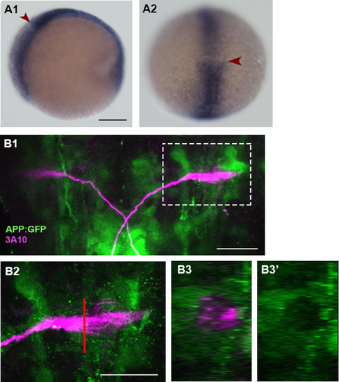
Appb is expressed at 10 hpf but not in the mature Mauthner cell. In situ hybridization of appb mRNA showed stronger expression in the hindbrain at 10 hpf (A1, A2). Embryos are shown in lateral (A1) and dorsal view (A2). Arrowhead indicates the boundary between hindbrain. Co-labeling of Mauthner cell with Tg(Appb:GFP) transgenic fish line (B1-B3′) at 24 hpf. (B2) Higher magnification of boxed region in B1. (B3-B3′) Single plane lateral view across the M-cell, indicated by red vertical line in B2. Scale bars: A1, 100 µm: B1, B2, 25 µm. |
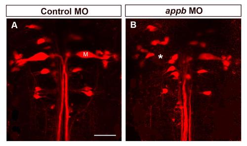
Knock down of Appb causes alteration in Mauthner cell formation. Dorsal view of the reticulospinal neurons in live zebrafish larvae at 4 dpf labeled with rhodamine dextran via retrograde labeling (A-B). The large M-cell is denoted by M (A). The position of the missing M-cell is noted by an asterisk (B). Scale bar, 50 µm. |
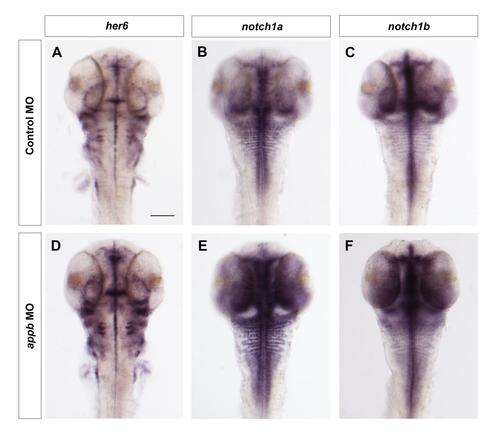
Increased Notch signaling in appb morphants. Dorsal view of the flat-mounted control MO (A-C) and appb MO (D-F) injected embryos (anterior at the top) at 48 hpf showing her6 (A, D), notch1a (B, E) and notch1b (C, F) expression levels in the hindbrain. Scale bar, 100 µm. |
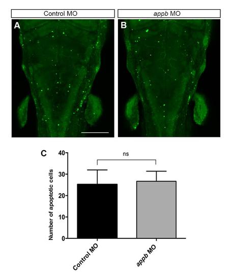
Cell death analysis in appb morphants. Acridine orange (AO)-labeled embryos at 48 hpf (A-B). Confocal images (anterior at the top) of hindbrain labeled with acridine orange (AO) in control MO (A) and appb MO-injected (B) embryos at 48 hpf. Bar graph displays the quantification of AO positive cells (C). Scale bar, 50 µm. ****p < 0.0001 |
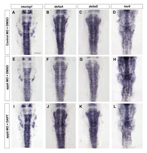
DAPT treatment rescues the expression of neurog1, deltaA/D and her6 in appb morphants. Dorsal view of flat-mounted control MO + DMSO (A-D) and appb MO + DMSO (E-H) and appb MO + DAPT treated embryos (anterior at the top) at 24 hpf showing neurog1 (A, E, I), deltaA (B, F, J), deltaD (C, G, K) and her6 (D, H, L) expression levels in the hindbrain. Scale bar, 75 µm. |
Reprinted from Developmental Biology, 413(1), Banote, R.K., Edling, M., Eliassen, F., Kettunen, P., Zetterberg, H., Abramsson, A., β-Amyloid precursor protein-b is essential for Mauthner cell development in the zebrafish in a Notch-dependent manner, 26-38, Copyright (2016) with permission from Elsevier. Full text @ Dev. Biol.