Fig. 2
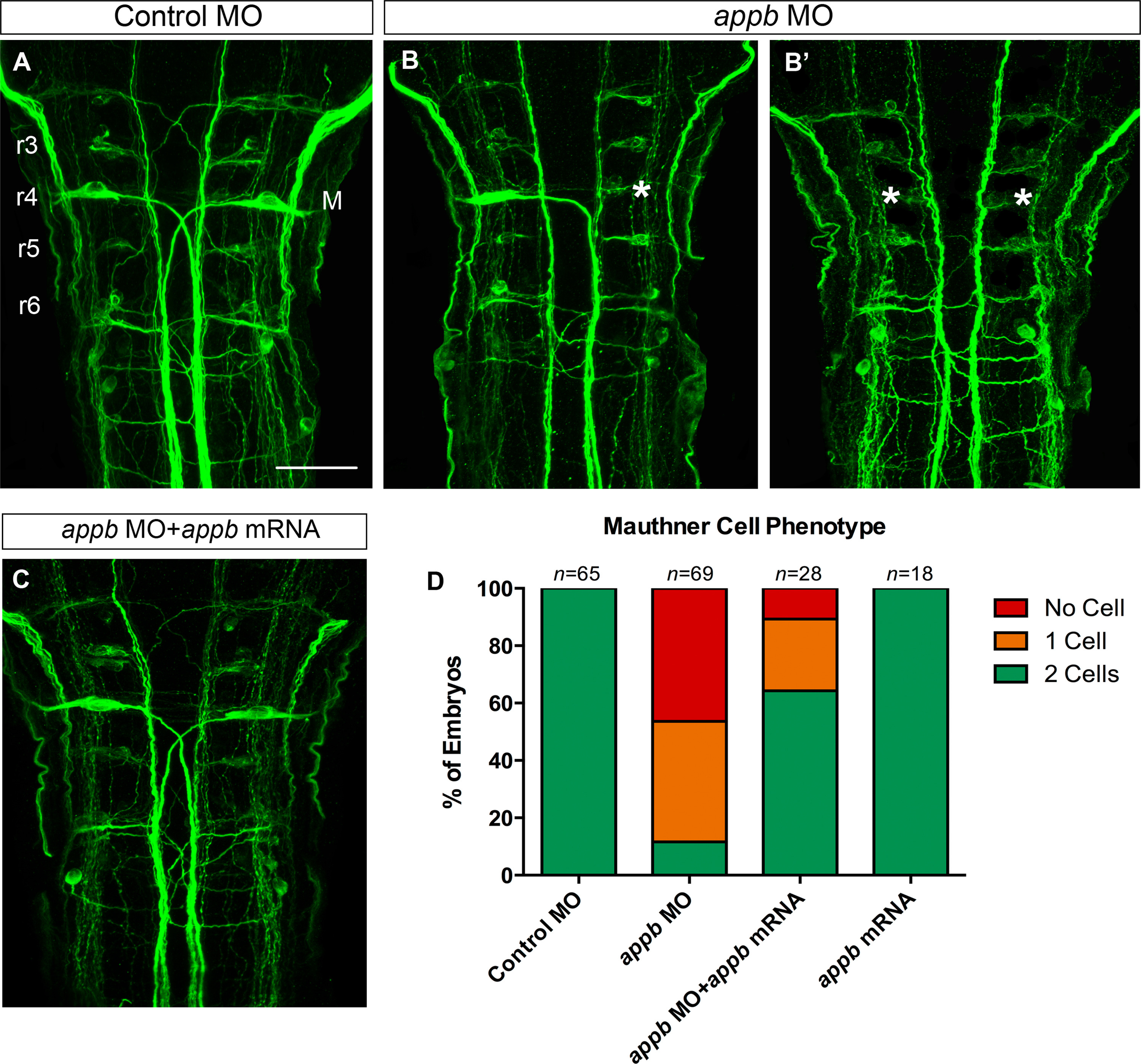
Knockdown of Appb inhibit Mauthner cell development. Whole-mount immunostaining of embryos at 48 hpf (A-C). Confocal images (anterior to the top) of hindbrain stained with anti-neurofilament RMO44 antibody marking reticulospinal neurons in control MO (A) and appb MO injected (B,B2) embryos. Note the 1-cell/no-cell phenotype in morphants. The large M-cells (M, in A) are located in r4. The absent M-cell is noted by an asterisk (B,B′). M-cell number is rescued in embryos coinjected with appb MO and appb mRNA (C). Cell counts of M-cell number (D). M, Mauthner cell; n, number of embryos and r3-r6, rhombomeres 3-6. Scale bar, 50 µm.
Reprinted from Developmental Biology, 413(1), Banote, R.K., Edling, M., Eliassen, F., Kettunen, P., Zetterberg, H., Abramsson, A., β-Amyloid precursor protein-b is essential for Mauthner cell development in the zebrafish in a Notch-dependent manner, 26-38, Copyright (2016) with permission from Elsevier. Full text @ Dev. Biol.

