
- Title
-
Bmp activity establishes a gradient of positional information throughout the entire neural plate
- Authors
- Barth, K.A., Kishimoto, Y., Rohr, K.B., Seydler, C., Schulte-Merker, S., and Wilson, S.W.
- Source
- Full text @ Development
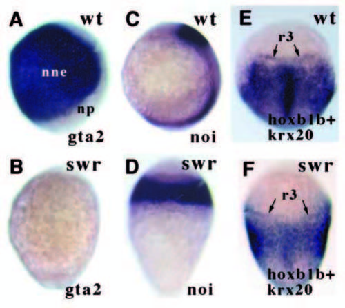
The ectoderm of swr- embryos is neuralised. In lateral views, dorsal is to the right; in dorsal views, anterior is up. (A,B) Lateral views of gta2 expression (prospective epidermis) in bud stage embryos. (C,D) Lateral views of noi/pax2.1 expression (prospective midbrain) in 1-2 somite stage embryos. (E-F) Dorsal views of hoxb1b (posterior tissue) and krx20 (r3 + r5) in 1-2 somite stage embryos. Scale in this and subsequent figures: the yolk ball of wildtype embryos is 700 μm across. nne, non-neural ectoderm; np, neural plate; r3, rhombomere 3; wt, wild-type. EXPRESSION / LABELING:
|
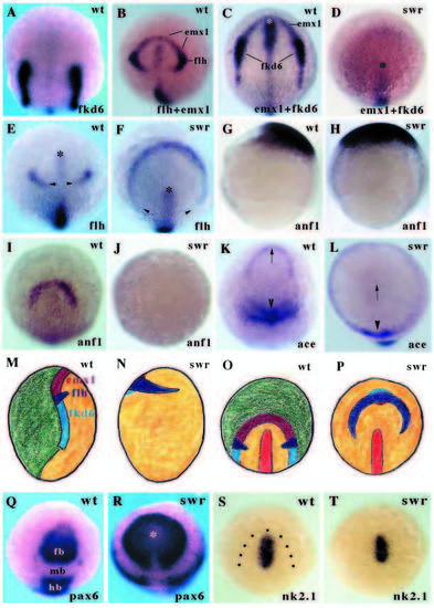
Swirl/Bmp2b affects fates at the margin of the neural plate at all AP levels. Animal pole (A-F,I-L,O-T) and lateral (G,H,M,N) views of 1-2 somite stage (except G,H) embryos. (A) fkd6 expression in prospective neural crest cells at the margins of the neural plate. (B) flh expression in prospective dorsal diencephalon and emx1 in prospective telencephalic cells around the rostral margin of the neural plate. The posterior limit of emx1 expression overlaps with flh expression. The posterior boundary of flh expression is at the anterior boundary of fkd6 expression. (C,D) emx1 and fkd6 expression. In the swr- embryo, marginal expression of both genes is absent. The asterisk indicates the rostral limit of axial tissue (also marked by transient fkd6 and flh expression in rostral midline) in this and other panels. (E-F) flh expression in prospective dorsal diencephalon. In the swr- embryo, flh expression is radially expanded into the ventral ectoderm and the gap between the domains of flh expression on the dorsal side of the embryo is expanded (arrowheads). (G-J) anf1 expression in 80% (G-H) and 1-2 somite stage (I-J) embryos. anf1 is initially expanded in swr- embryos but is lost by the stage that expression is restricted to prospective telencephalic cells. (K,L) ace/fgf8 expression. Telencephalic expression (arrow) is absent in swr- embryos whereas more caudal expression domains (arrowheads) are radialised. (M-P) Schematic illustrations of marginal neural plate cell fates in wild-type (M,O) and swr- (N,P) embryos. M,N are lateral views and O,P animal pole views of bud-1 somite stage embryos. Green is nonneural tissue, yellow is neural plate. Pale blue shows fkd6 in prospective neural crest, dark blue shows flh expression in dorsal diencephalic cells and maroon represents emx1/anf/ace expression in prospective telencephalic cells. Marginal cell fates are absent in anterior and posterior regions but at the level of the diencephalon, flh is expanded and some fkd6 and emx1 expression may be retained. (Q,R) pax6 expression. Diencephalic and hindbrain pax6 expression is expanded in the mutant embryo. The asterisk indicates the anterior limit of the axis. (S,T) nk2.1 expression in the prospective hypothalamus of wild-type and swr- embryos. Dots indicate the approximate margin of the neural plate. fb, forebrain; hb, hindbrain; mb, midbrain. EXPRESSION / LABELING:
|
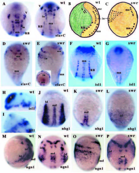
Swirl/Bmp2b affects neurogenesis throughout the neural plate. All embryos are 1-2 somite stage (except H,I which are 14 somite stage). (A) Animal pole (left) and dorsal (right) views of elavC expression in neurons in the neural plate of wildtype embryos. (B,C) Schematic illustrations of lateral views of neuronal patterning in wild-type and swr- embryos. Green is non-neural ectoderm, yellow is the neural plate and the dots represent neurons. In swr- embryos, trigeminal and Rohon-Beard neurons are absent while interneurons are massively radially expanded on the ventral side of the embryo. (D) Dorsal and (E) lateral views of elavC expression in swr- embryos. The interneuron population is hugely expanded. The inset shows an animal pole view of the same embryo. (F,G) Dorsal views of isl1 expression in wild-type and swr- embryos. The Rohon-Beard and trigeminal neurons are absent in the swr- mutant. (H,I) isl1 expression in wild-type and swr- embryos. Neurogenesis is considerably reduced in the anteriormost CNS of the swr- embryo as compared to wild type. (J,K) Dorsal and (L) ventral views of nhg1 expression in the ectoderm of wild-type and swr- embryos. The lateral stripes of nhg1 expression are either absent (K) or are expanded and fused on the ventral side in swr- embryos (L). (M,O) Lateral and (N,P) dorsal views of ngn1 in wild-type and swr- embryos. In the swr- embryo, lateral domains of expression are lost, intermediate domains hugely expanded and expression in the vicinity of motorneurons is also slightly broader. in, interneurons; id, intermediate domain; ld, lateral domain; me, medial neurons; md, medial domain; mn, motor neurons; RB, Rohon- Beard neurons; V, trigeminal neurons. EXPRESSION / LABELING:
PHENOTYPE:
|
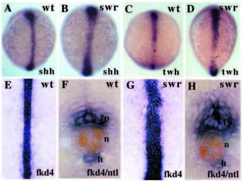
Floorplate tissue is expanded in swr- embryos. (A-D) Dorsal views of shh (A,B, 1-2 somite stage) and twh (C,D, 3 somite stage) expression in axial tissue. shh is expressed in ectodermal and mesodermal tissue whereas by the 3 somite stage, twh is limited to ectodermal tissue (Ekker et al., 1995). The expression of both genes is slightly expanded in swr- embryos. (E-H) Whole-mount views and transverse sections of fkd4 expression in floorplate and hypochord and Ntl (Schulte-Merker et al., 1994) expression in the notochord in 15 somite stage embryos. Floorplate fkd4 expression is expanded in the swr- mutants. fp, floorplate; h, hypochord; n, notochord. EXPRESSION / LABELING:
|

Noggin-injected embryos are more severely dorsalised than swr- embryos. Lateral (A), animal pole (B-D) and dorsal (E-H) views of of 1-2 somite stage (except A, 90% epiboly) embryos injected with noggin RNA. (A) anf and noi expression is radialised in noggin-injected embryos (similar to swr- embryos; compare with Figs 1D and 2H). (B) nk2.1 expression is retained in the prospective hypothalamus of the injected embryo. (C,D) Marginal neural plate expression of flh, emx1 and fkd6 is lost in noggin-injected embryos (compare with Fig. 2D,F). (E) Mild and (F) massive expansion of isl1 expressing motor neurons in noggin-injected embryos. The asteriks indicates the pillow. (G) Radial expansion of elavC expressing medial neurons in a severely dorsalised noggin-injected embryo. (H) shh expression in axial tissue in a noggin-injected embryo. me, medial neurons; mn, motor neurons. |
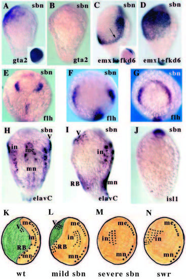
Sbn affects marginal cells fates and patterns of neurogenesis. All panels show 1-2 somite stage embryos. (A,B) Lateral views of gta2 expression (prospective epidermis) in mild (A) and severe (B) sbn- embryos. The inset panel shows expression in a wild-type embryo. Expression is retained in the anterior ventral region of the mildly affected embryo. (C,D) Lateral views of emx1 (prospective telencephalon) and fkd6 (prospective neural crest) expression in mild (C) and severe (D) sbn- embryos. The inset panel shows expression in a wild-type embryo. In the mildly affected embryo, expression is relatively normal in rostral regions, consistent with retention of gta2 expression (see A) in ventral ectoderm. However, more caudally, fkd6 expression is radialised (arrow). In the severely affected embryo, emx1 and fkd6 are expanded throughout the ventral ectoderm consistent with loss of gta2 in this region (see B). (E-G) Animal pole views of flh expression (prospective epiphysis) in progressively more severe sbn- embryos. flh expression is variably expanded in the sbn- embryos (see Fig. 2E,F for wild-type and swr- expression). (H) Dorsal and (I) lateral views of elavC expression in a sbn- embryo. Wild-type pattern of expression is shown in Fig. 3A,B. In these mildly affected sbn- embryos, interneurons are slightly expanded and dorsal sensory neurons are broadly expanded on the ventral side of the embryo. (J) Ventral view of isl1 expression in a severely affected sbn- embryo. Trigeminal and Rohon-Beard neurons are absent. (K-N) Summary schematics of patterns of neurogenesis in wild-type, sbn- and swr- embryos. In the mildly affected sbn- embryo, some non-neural ectoderm is retained and trigeminal and RB neurons are expanded on the ventral side of the embryo. In the severe sbn- embryo, trigeminal and RB neurons are lost and in the swr- embryo, interneurons are expanded throughout the ventral ectoderm. in, interneurons; me, medial neurons; mn, motor neurons; RB, Rohon-Beard neurons; V, trigeminal neurons. EXPRESSION / LABELING:
PHENOTYPE:
|