
IMAGE
Fig. 1
- ID
- ZDB-IMAGE-140317-26
- Genes
- Publication
- Barth et al., 1999 - Bmp activity establishes a gradient of positional information throughout the entire neural plate
- All Figures
- Figures for Barth et al., 1999
Image
|
Figure Caption
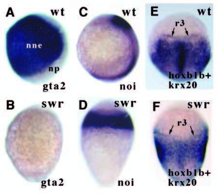
Fig. 1 The ectoderm of swr- embryos is neuralised. In lateral views, dorsal is to the right; in dorsal views, anterior is up. (A,B) Lateral views of gta2 expression (prospective epidermis) in bud stage embryos. (C,D) Lateral views of noi/pax2.1 expression (prospective midbrain) in 1-2 somite stage embryos. (E-F) Dorsal views of hoxb1b (posterior tissue) and krx20 (r3 + r5) in 1-2 somite stage embryos. Scale in this and subsequent figures: the yolk ball of wildtype embryos is 700 μm across. nne, non-neural ectoderm; np, neural plate; r3, rhombomere 3; wt, wild-type.
Figure Data
Acknowledgments
This image is the copyrighted work of the attributed author or publisher, and
ZFIN has permission only to display this image to its users.
Additional permissions should be obtained from the applicable author or publisher of the image.
Full text @ Development