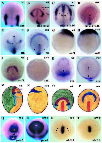
Swirl/Bmp2b affects fates at the margin of the neural plate at all AP levels. Animal pole (A-F,I-L,O-T) and lateral (G,H,M,N) views of 1-2 somite stage (except G,H) embryos. (A) fkd6 expression in prospective neural crest cells at the margins of the neural plate. (B) flh expression in prospective dorsal diencephalon and emx1 in prospective telencephalic cells around the rostral margin of the neural plate. The posterior limit of emx1 expression overlaps with flh expression. The posterior boundary of flh expression is at the anterior boundary of fkd6 expression. (C,D) emx1 and fkd6 expression. In the swr- embryo, marginal expression of both genes is absent. The asterisk indicates the rostral limit of axial tissue (also marked by transient fkd6 and flh expression in rostral midline) in this and other panels. (E-F) flh expression in prospective dorsal diencephalon. In the swr- embryo, flh expression is radially expanded into the ventral ectoderm and the gap between the domains of flh expression on the dorsal side of the embryo is expanded (arrowheads). (G-J) anf1 expression in 80% (G-H) and 1-2 somite stage (I-J) embryos. anf1 is initially expanded in swr- embryos but is lost by the stage that expression is restricted to prospective telencephalic cells. (K,L) ace/fgf8 expression. Telencephalic expression (arrow) is absent in swr- embryos whereas more caudal expression domains (arrowheads) are radialised. (M-P) Schematic illustrations of marginal neural plate cell fates in wild-type (M,O) and swr- (N,P) embryos. M,N are lateral views and O,P animal pole views of bud-1 somite stage embryos. Green is nonneural tissue, yellow is neural plate. Pale blue shows fkd6 in prospective neural crest, dark blue shows flh expression in dorsal diencephalic cells and maroon represents emx1/anf/ace expression in prospective telencephalic cells. Marginal cell fates are absent in anterior and posterior regions but at the level of the diencephalon, flh is expanded and some fkd6 and emx1 expression may be retained. (Q,R) pax6 expression. Diencephalic and hindbrain pax6 expression is expanded in the mutant embryo. The asterisk indicates the anterior limit of the axis. (S,T) nk2.1 expression in the prospective hypothalamus of wild-type and swr- embryos. Dots indicate the approximate margin of the neural plate. fb, forebrain; hb, hindbrain; mb, midbrain.
|
