
- Title
-
Insulin-like growth factor-2 regulates early neural and cardiovascular system development in zebrafish embryos
- Authors
- Hartnett, L., Glynn, C., Nolan, C.M., Grealy, M., and Byrnes, L.
- Source
- Full text @ Int. J. Dev. Biol.
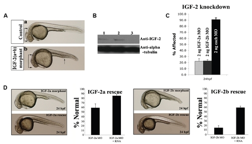
IGF-2 morpholinos efficiently and specifically knockdown IGF-2 in vivo. (A) Phenotype of (a) control injected and (b) IGF-2(a+b) morphant embryo at 24 hpf. IGF-2(a+b) morphant embryos are ventralised with a shorter body, disrupted brain structures (double arrows), reduced eyes, disrupted somites and an expanded intermediate cell mass (arrow). (B) Western blot showing IGF-2 and alpha-tubulin at 24 hpf in (1) uninjected control embryos, (2) embryos injected with control morpholinos or (3) embryos injected with IGF-2(a+b) morpholinos. (C) Igf-2a and igf-2b mediate their effects on development in a synergistic fashion. (D) Co-injection of igf-2a or igf-2b RNA with the corresponding morpholino rescues the IGF-2 morphant phenotype. Rescued embryos display normal body size, eyes and brain structures at 24 hpf. Results shown are a summary of three independent experiments (n ≥ 80). Error bars indicate the standard deviation of the mean. PHENOTYPE:
|
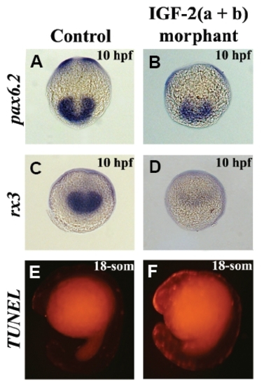
IGF-2 is required for the development of anterior neural structures during gastrulation and plays an anti-apoptotic role during segmentation. (A,C) Control morpholino injected embryos show normal pax6.2 and rx3 expression. (B,D) IGF-2(a+b) morphant embryos show reductions in pax6.2 and rx3 expression. (E) Embryos injected with control morpholinos show low levels of apoptosis during zebrafish segmentation. (F) Knockdown of igf-2a and igf-2b results in an increase in apoptosis in the anterior region of the embryo and in the developing spinal cord. Frequency of embryos displaying this staining pattern; B, 14/ 32; D, 22/40; F, 17/28. (A-D) are views of the future anterior region; (E,F) are lateral views. |
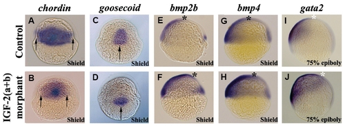
IGF-2 regulates the expression of genes involved in dorsal-ventral patterning. (A,C) Embryos injected with control morpholinos show normal chordin and goosecoid expression. (B,D) Expression of chordin and goosecoid is reduced in IGF-2(a+b) morphant embryos (arrows). (E,G,I) Embryos injected with control morpholinos show normal bmp2b, bmp4 and gata2 expression. (F,H,J) Expression of bmp2b, bmp4 and gata2 is expanded towards the dorsal side in IGF-2(a+b) morphant embryos (asterisks). Frequency of embryos displaying this staining pattern; B, 13/26; D, 17/31; F, 16/25; H, 18/32; J, 26/34. All embryos are shown in a lateral view. EXPRESSION / LABELING:
PHENOTYPE:
|

Blood circulation is disrupted when IGF signalling is reduced. (A) Control morpholino injected embryos stained with O-dianisidine at 72 hpf show normal blood circulation. (B) IGF-2(a+b) morphant embryo with reduced circulating blood. Grey arrows indicate blood in the heart region, red arrows blood circulation in the intersomitic vesicles and grey arrowheads the intermediate cell mass. (C,F) Embryos injected with control morpholinos showing normal scl and gata1 expression at 26 hpf. (D,G) Knockdown of igf-2a and igf-2b results in an increase in scl and gata1 expression and an expansion of expression outside the intermediate cell mass. (E,H) DN-IGF-1R injected embryos show an increase in scl and gata1 expression. Arrowhead indicates position of intermediate cell mass and arrows indicate extent of expression along the embryo. Frequency of embryos displaying this staining pattern; B, 73/86; D, 37/48; E, 30/30; G, 48/58; H, 26/29. All embryos are shown in lateral view. EXPRESSION / LABELING:
PHENOTYPE:
|
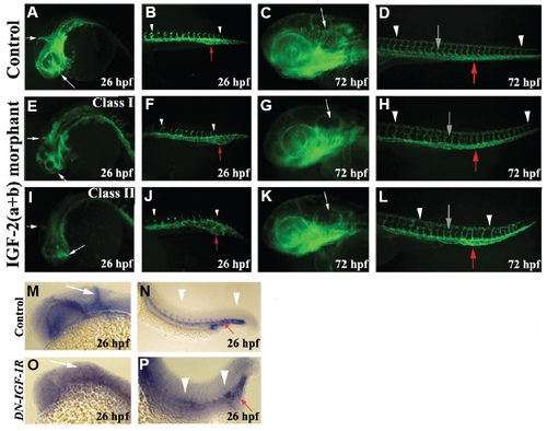
Angiogenesis is compromised when IGF signalling is reduced. (AD) fli1:EGFP transgenic embryos injected with control morpholinos showing normal vascular development. (E-H) Class I IGF-2 morphant embryo. The intensity of GFP expressing cells in the head and eyes is reduced while the basic pattern of vasculature remains intact. The intermediate cell mass is mildly expanded and the intersomitic vessels are reduced. By 72 hpf, the parachordal vessel is incompletely formed. (I-L) Class II IGF-2 morphant embryo. The sprouting of vessels in the head and eyes are reduced. Intersomitic vessel sprouting is irregular and reduced, the intermediate cell mass is expanded and the parachordal vessel is disrupted. (M,N) Control embryo at 26 hpf showing normal expression of flk1 in the vasculature. (O,P) Angiogenesis is disrupted in DN-IGF-1R injected embryos as shown by flk1 expression (n=14/29). White arrows indicate the vasculature in the head and eyes. White arrowheads point to the intersomitic vessels, red arrow points to the intermediate cell mass and the grey arrow indicates the parachordal vessel. All embryos are shown in a lateral view. PHENOTYPE:
|

IGF signalling is critical for cardiac valve development in zebrafish. (A,C) Heart region of IGF-2a or IGF- 2b morphant embryos at 72 hpf. Hearts are enlarged and incompletely looped (white outline) with blood reflux between the two chambers. (B,D) Co-injection of igf-2a or igf-2b RNA with the corresponding morpholino rescues the IGF-2 morphant phenotype. Hearts in rescued embryos are identical to hearts in uninjected embryos. (E,J) Expression of bmp4 and notch1b in embryos injected with control morpholinos. Note the restriction of expression at the atrioventricular boundary (black arrowheads). The white outline indicates the shape of the heart. (F,K) Bmp4 and notch1b are restricted to the atrioventricular boundary in IGF- 2a morphant embryos. (G,H,L,M) IGF-2b and IGF-2(a+b) morphant embryos show a loss of bmp4 and notch1b expression at the atrioventricular boundary (black arrowheads). The heart is also incompletely looped at this stage (white outline). (I,N) DN-IGF-1R injected embryos show a loss of bmp4 and notch1b at the atrioventricular boundary. (O) Expression of eln2 at the outflow tract of the heart in embryos injected with control morpholinos (white arrowhead). (P,Q,R) IGF-2a, IGF-2b and IGF-2(a+b) morphant embryos show a decrease of eln2 expression at the outflow tract. (S) DN-IGF-1R injected embryos show a loss of eln2 at the outflow tract. Frequency of embryos displaying this expression pattern: F, 30/30; G, 20/40; H, 15/27; I, 29/ 44; K, 33/33; L, 14/32; M, 12/30; N, 49/56; P, 5/24; Q, 31/36; R, 24/26; S, 31/39. (T) Normal cardiac looping in control injected embryos at 72 hpf as shown by the expression of cmlc2 (heart loops to the right in this view). EXPRESSION / LABELING:
PHENOTYPE:
|
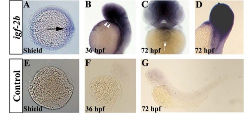
Expression of igf-2b during zebrafish embryogenesis. Whole mount in situ hybridisation was performed using igf-2b-specific antisense and sense probes. (A) igf-2b is expressed in the zebrafish embryonic shield (black arrow). (B) igf-2b is expressed in the developing anterior neural structures and the heart (white arrowheads). (C, D) By 72 hpf, igf-2b is expressed in the heart and in the anterior region of the embryo. (E, F, G) Embryos hybridised to the sense igf-2b probe. Frequency of embryos displaying this staining pattern; A, 51/55; B, 31/31; C, D, 26/26; E, 50/50; F, 28/28; G, 31/31. A, animal pole view of the embryo with dorsal to the right. B, D, E, F, G are shown in lateral view and C is shown in ventral view. EXPRESSION / LABELING:
|

Expression of fgf8 and bozozok in IGF-2 morphant embryos. (A-D) Knockdown of igf-2a and/or igf-2b does not disrupt patterning of the telencephalon (arrowhead) and the midbrain-hindbrain boundary (asterisk) during the segmentation stage in zebrafish development. (Inset) Anterior region of 24 hpf control and morphant embryos showing disrupted neural structures in morphants. (E-H) Expression of bozozok is unaffected in IGF-2 morphant embryos. Frequency of embryos displaying this staining pattern; B, 30/30; C, 28/28; D, 32/32; F, 60/60; G, 59/59; H, 42/42. All embryos are shown in a lateral view. EXPRESSION / LABELING:
|
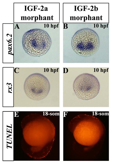
IGF-2a and IGF-2b are required for the development of anterior neural structures during zebrafish gastrulation and play anti-apoptotic roles during segmentation. (A, B) Knockdown of igf-2a or igf-2b results in a reduction of pax6.2 expression in the developing eye and midline region. (C, D) Knockdown of igf-2a or igf-2b causes a reduction of rx3 expression. (E) Knockdown of igf-2a results in an increase in apoptosis, particularly in the developing spinal cord. (F) Knockdown of igf-2b results in an increase in apoptosis, particularly in the anterior region of the embryo. Frequency of embryos displaying this staining pattern; A, 14/32; B, 8/20; C, 15/39; D, 14/33; E, 17/30; F, 12/33. A-D are views of the future anterior region and E, F are lateral views. EXPRESSION / LABELING:
PHENOTYPE:
|
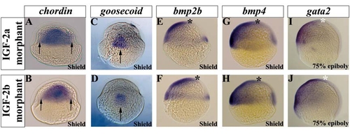
IGF-2a and IGF-2b regulates the expression of genes involved in dorsal-ventral patterning. (A, B) Expression of chordin is reduced in IGF-2a and IGF-2b morphant embryos. Arrows indicate the width of the chordin expression domain. (C, D) Expression of goosecoid is reduced in IGF-2a and IGF-2b morphant embryos (arrows). (E-J) Expression of bmp2b, bmp4 and gata2 is expanded towards the dorsal side of IGF-2a and IGF-2b morphant embryos (asterisks). Frequency of embryos displaying this staining pattern; A, 13/27; B, 17/35; C, 13/39; D, 17/31; E, 13/27; F, 16/32; G, 12/28; H, 15/36; I, 17/33; J, 21/39. All embryos are shown in a lateral view. EXPRESSION / LABELING:
PHENOTYPE:
|
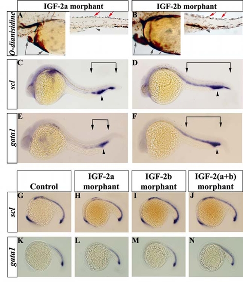
Defects in blood development are coincident with the onset of circulation in IGF-2 morphant embryos. (A, B) O-dianisidine stained IGF- 2a and IGF-2b morphant embryos with reduced circulating blood at 72 hpf (heart and tail regions). Grey arrows indicate blood pooling under the heart, red arrows loss of blood circulation in the intersomitic vesicles and grey arrowheads the intermediate cell mass. (C, E) IGF-2a morphant embryos show an increase in scl and gata1 expression at the intermediate cell mass. (D, F) Knockdown of igf-2b results in an increase in the region of scl and gata1 and expression is expanded outside the intermediate cell mass. Arrowhead indicates position of intermediate cell mass and arrows indicate extent of expression along the embryo. (G, K) 22-somite stage embryos injected with control morpholino(s) showing normal scl and gata1 expression. (H-J, L-N) Expression of scl and gata1 is unaffected in IGF-2 morphant embryos before circulation commences. Frequency of embryos displaying this expression pattern: A, 20/64; B, 39/47; C, 12/43; D, 37/51; E, 19/75; F, 73/84; H, 32/32; I, 29/29; J, 35/35; L, 23/25; M, 29/31; N, 33/33. All embryos are shown in a lateral view. EXPRESSION / LABELING:
PHENOTYPE:
|
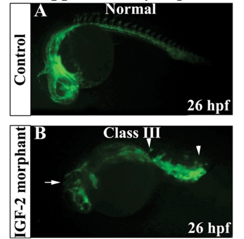
Vasculogenesis is unaffected in IGF-2 morphant embryos. (A) Control fli1:EGFP transgenic embryo displaying normal vascular development at 26 hpf. (B) Class III IGF-2 morphant embryo displaying severe effects on angiogenesis, however the basic vasculature is laid down. PHENOTYPE:
|

Unillustrated author statements PHENOTYPE:
|