
Fig. S1
- ID
- ZDB-IMAGE-100504-47
- Genes
- Publication
- Hartnett et al., 2010 - Insulin-like growth factor-2 regulates early neural and cardiovascular system development in zebrafish embryos
- All Figures
- Figures for Hartnett et al., 2010
|
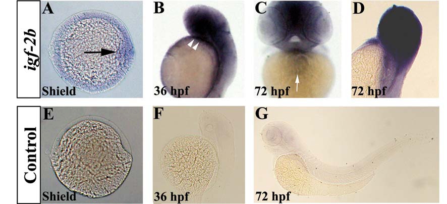
Fig. S1 Expression of igf-2b during zebrafish embryogenesis. Whole mount in situ hybridisation was performed using igf-2b-specific antisense and sense probes. (A) igf-2b is expressed in the zebrafish embryonic shield (black arrow). (B) igf-2b is expressed in the developing anterior neural structures and the heart (white arrowheads). (C, D) By 72 hpf, igf-2b is expressed in the heart and in the anterior region of the embryo. (E, F, G) Embryos hybridised to the sense igf-2b probe. Frequency of embryos displaying this staining pattern; A, 51/55; B, 31/31; C, D, 26/26; E, 50/50; F, 28/28; G, 31/31. A, animal pole view of the embryo with dorsal to the right. B, D, E, F, G are shown in lateral view and C is shown in ventral view.