
- Title
-
Proteomics of early zebrafish embryos
- Authors
- Link, V., Shevchenko, A., and Heisenberg, C.P.
- Source
- Full text @ BMC Dev. Biol.
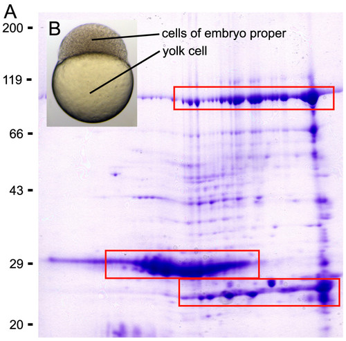
The bulk of total protein in the early embryo is yolk protein. A. Coomassie blue stained 2D gel (pI 3-10) of 1 mg protein extracted from shield stage embryos (6 hpf) without prior removal of the yolk. Several isoforms and degradation products of the predominant yolk protein Vitellogenin were spread over large parts of the gel (three boxes). Vitellogenin was identified by mass spectrometry. B. Embryo at high stage (3 1/3 hpf). The volume of the yolk cell exceeds the volume of the cells constituting the embryo proper. |

Improved Western blotting results. Embryos at high stage (3 1/3 hpf), 50% epiboly (5 1/4 hpf), 70% epiboly (7 hpf) and tailbud stage (10 hpf) were deyolked, separated by SDS-gel electrophoresis and Coomassie stained (A) or blotted and immunodetected with antibodies against Tubulin (55 kD) and Moesin (78/80 kD apparent molecular weight) (B). Note that total protein amount was lower in deyolked samples, therefore more embryos could be loaded per lane: 1 embryo with yolk (Y), 15 embryos deyolked (D), 15 embryos deyolked and washed twice (W). Consequently, signal intensities of cellular proteins were increased. |
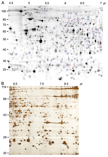
2D gels. 2D gel electrophoresis of samples derived from embryos at 80% epiboly (8 hpf) in the acidic as well as in the basic range. A. pI 4-7 (24 cm), fluorescently labelled with Cy2, 50 μg protein/dye. Spots processed for MS-identification are labelled (ID of Additional file 1). B. pI 6-9 (18 cm), silver stain. EXPRESSION / LABELING:
|