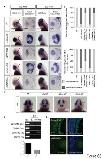
(A) WISH assays monitoring the expression of vsx2 at 48 hpf or otx2 at 72 hpf in lateral views or retinal cross-sections from WT, znf644a morphant or znf644b morphant embryos, as well as morphant embryos rescued by co-injection of the indicated mRNA. Red arrows indicate hindbrain neural cell populations that express vsx2. (B) Frequency at which the indicated embryos exhibited normal or mislocalized expression of vsx2 (top) or otx2 (bottom). (C) WISH assays monitoring the expression of lef1 at 48 hpf (dorsal views) in WT, g9a morphant, znf644a morphant, or znf644b morphant embryos. Red arrows point to lef1-positive midbrain regions, and red arrowheads point to lef1-expressing cells of the anterior hypothalamus. (D) ChIP-PCR assays monitoring the levels of H3K9me2 at the indicated positions near the TSS of vsx2 or ccnd1 genes (as in Figure 3C) at 48 hpf. An anti-H3K9me2 antibody and an isotype control (IgG) antibody were used for ChIP, followed by PCR amplification (35 cycles). Densitometry analysis revealed a ~50% reduction in the levels of H3K9me2 at the ccnd1 promoter, and complete loss of H3K9me2 levels at the vsx2 promoter in g9a morphant embryos. (E) Immunostaining with H3K9me2 antibody demonstrates a global reduction in nuclear expression in the g9a morphant retinas at 48 hpf compared to controls.
|
