
Fig. 6
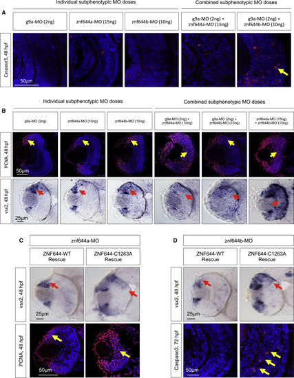
The Retinal Functions of znf644a and znf644b Are Dependent on Functional and Physical Interactions with g9a (A) Immunostaining of cleaved Caspase3 (red) in retinal cross-sections of embryos injected with individual or combined subphenotypic doses of g9a-MO, znf644a-MO, or znf644b-MO (n = 3 in each group). Yellow arrows highlight apoptotic cells. (B) (Top) Immunostaining of PCNA (n = 6 in each group) in retinal cross-sections from embryos injected with individual and combined subphenotypic doses of MOs (n = 6 in each group). Yellow arrows denote the position of the PCNA+ population. (Bottom) WISH assays monitoring the expression of vsx2 in retinal cross-sections of embryos injected with individual subphenotypic doses (n = 10 in each group) and combined subphenotypic co-injection of g9a-MO and znf644a-MO (n = 10), g9a-MO and znf644b-MO (n = 13), and znf644a-MO and znf644b-MO (n = 10). Red arrows denote the position of the vsx2 expressing cells. (C) (Top) WISH assays in retinal cross-sections monitoring vsx2 expression at 48 hpf in znf644a morphant embryos rescued by co-injection of either WT human ZNF644 mRNA (n = 12) or C1263A mutant mRNA (n = 9). (Bottom) Immunostaining of retinal cross-sections monitoring PCNA expression at 48 hpf showing rescue of znf644b morphants by co-injection of WT human ZNF644 mRNA (n = 3) but not C1263A mutants (n = 3). (D) (Top) WISH assays in retinal cross-sections at 48 hpf monitoring vsx2 expression from znf644b morphant embryos rescued by co-injection of human WT ZNF644 mRNA (n = 14) or C1263A ZNF644 mutant mRNA (n = 9). (Bottom) Immunostaining of retinal cross-sections monitoring cleaved Caspase3 at 72 hpf in znf644b morphant embryos rescued by co-injection of WT human ZNF644 mRNA (n = 3) or a C1263A mutant (n = 3). Red arrows highlight vsx2 expression domains, and yellow arrows highlight PCNA+ cell populations (C) or apoptotic cells (D). |
| Gene: | |
|---|---|
| Antibodies: | |
| Fish: | |
| Knockdown Reagents: | |
| Anatomical Terms: | |
| Stage: | Long-pec |
| Fish: | |
|---|---|
| Knockdown Reagents: | |
| Observed In: | |
| Stage: | Long-pec |