- Title
-
Fibroblast growth factor signaling is required for early somatic gonad development in zebrafish
- Authors
- Leerberg, D.M., Sano, K., Draper, B.W.
- Source
- Full text @ PLoS Genet.
|
Larval and juvenile fgf24 mutants have severely underdeveloped gonads and consequently develop as male. (A-C') fgf24 mutants are phenotypically male. A wild-type (WT) female has a distended belly (A) and protruding cloaca (arrow, A') compared to the slender belly and obscured cloaca of the wild-type (B, B') and mutant (C, C') males. Note the absence of pectoral fin in the fgf24 mutant (C) compared to wild-type animals (arrowheads, A, B). (D) Box and whisker plot representing the number of germ cells per wild-type and fgf24 mutant fish during larval development. Each dot represents the number of germ cells in one animal (Unpaired two-tailed t-test; n = 15 for both 8 dpf wild-type and mutant, n = 13 for 10 dpf wild-type, n = 12 for 10 dpf mutant, n = 10 for 12 dpf wild-type, n = 11 for 12 dpf mutant, n = 16 for 14 dpf wild-type, n = 12 for 14 dpf mutant; * = P < .05; n.s. = no significance). (E-H') Representative confocal images of 12 dpf (E-F) and 40 dpf (G-H') wild-type (E, G-G') and fgf24 mutant (F, H-H') gonads. At 12 dpf, gonads of fgf24 mutants (F) have fewer Vasa positive germ cells (green) compared to wild-type (E). By 40 dpf, wild-type testes have many Tg(ziwi:EGFP) positive germ cells (green), both premeiotic and spermatogenic, that are enclosed by Tg(gsdf:mCherry) positive Sertoli cells (red) (G-G'). In contrast, gonads of 40 dpf fgf24 mutants are unorganized, lack Tg(gsdf:mCherry) expressing Sertoli cells, and contain few germ cells, all of which are premeiotic (H-H'). G' and H' are magnified views G and H, respectively. The boxed nuclei in G' and H' are magnified in the respective insets, with an arrow indicating the large nucleoli (DAPI only, in grey). E-H' are sagittal optical sections with anterior to the left. Nuclei labeled with DAPI (blue). Scale bars = 20 μm for E, F, G, H; 100 μm for G', H'. EXPRESSION / LABELING:
|
|
fgf24 is expressed in somatic gonad cells during wild-type larval development. (A, B, C, D, E) Single plane confocal micrographs of whole-mount larval gonads after fluorescent in situ hybridization detecting fgf24 (red). At 5 dpf, fgf24 is not detected by fluorescent in situ hybridization (A). By 8 dpf, fgf24 is detected in SGCs (B) and is restricted to an outer layer of SGCs at 10 (C) and 16 (D) dpf. By 20 dpf, expression is primarily restricted to SGCs on the dorsal edge of the gonad (E). (A', B', C', D', E') Gonads dissected from fgf24 mutants age-matched to the wild-type animals depicted in A, B, C, D, and E. Gonads are outlined with white dotted lines. A-E' are sagittal optical sections where anterior is to the left and dorsal is up. Nuclei labeled with DAPI (blue), germ cells labeled with Vasa (green). Scale bars = 20 μm. EXPRESSION / LABELING:
|
|
A second, inner layer of somatic gonad cells responds to Fgf24 signaling. (A-D, E) Single plane view of whole mount gonads after fluorescent in situ hybridization. (A-B) At 8 dpf, ~85% of wild-type (WT) fish express fgf24 (red) in SGCs, whereas ~56% express etv4 (green) (A). (C) etv4 is undetected in fgf24 mutant gonads (n = 14). (D) By 10 dpf, SGCs located externally express fgf24, whereas an inner layer of SGCs expresses etv4. (D') An orthogonal view of a Z stack reconstruction of the gonad in D (white line). (E) In 10 dpf fgf24 mutant gonads, etv4 is expressed at low levels or not at all. Germ cells labeled with Vasa (blue). Gonads are outlined with white dotted lines in A-C. (F-G) Antibody staining of 10 dpf gonads detecting phosphorylated Erk (pErk, red). Nuclei labeled with DAPI (blue), germ cells labeled with Vasa (green). SGCs of wild-type gonads (F), but not fgf24 mutant gonads (G), show Erk phosphorylation. (F', G') are pErk channel only of (F) and (G), respectively. A-D, E-G' are sagittal optical sections with anterior to the left. Scale bars = 20 μm. |
|
Fgf24 regulates gene expression of somatic gonad cells. (A-J) Single plane confocal micrographs of whole-mount larval gonads after fluorescent in situ hybridization detecting SGC markers (red). Nuclei labeled with DAPI (blue), germ cells labeled with Vasa (green). gata4 (A), nr5a1a (C), wt1a (E), cyp19a1a (G), and amh (I) are expressed in somatic gonad cells (SGCs) of wild-type larval gonads, whereas only wt1a can be detected in SGCs of fgf24 mutants (F). (K-M) Double fluorescent in situ hybridization and Vasa antibody staining (blue) of 12 dpf wild-type gonads (outlined by white dotted line). etv4, cyp19a1a, and amh are expressed in non-overlapping SGC domains (n = 12, 12, and 3 for K, L, and M, respectively). In E, arrows denote inner SGCs; arrowheads denote outer SGCs. A-M are sagittal optical sections with anterior to the left. Scale bars = 20 μm. |
|
fgf24 mutant somatic gonad cells and germ cells have low proliferation. (A-C') 10 dpf wild-type (WT; n = 7; A, A') and fgf24 mutant (n = 7; C, C') gonads exhibit rare Cc3 staining (red). Wild-type and fgf24 mutant animals have similar numbers of Cc3(+) germ cells (mean ± SEM = 0.14 ± 0.14, 0, respectively; P = 0.36) and Cc3(+) SGCs (mean ± SEM = 0.57 ± 0.3, 0.43 ± 0.2, respectively; P = 0.7). (B, B') In a 10 dpf wild-type gonad, one Tg(ziwi:EGFP) positive germ cell (green) stains positive for Cc3 (red). (D-F) EdU incorporation from 8 to 9 dpf. Fish exposed to 200 μM EdU between 8 and 9 dpf were euthanized and stained for Vasa (green) and EdU (red). A lower percentage of mutant somatic gonad cells have incorporated EdU compared to wild-type (mean = 41% and 70%, respectively; P < .001; arrow = EdU(+) SGC, arrowhead = EdU(-) SGC). (G-I) Antibody staining against phospho-Histone H3 (pHH3, red) and Vasa (green). At 8 dpf, germ cells of fgf24 mutants are equally likely to be pHH3(+) as those of wild-type animals (mean = 22% and 16%, respectively; P > 0.05), however by 10 dpf, mutants have a significantly lower proportion of pHH3(+) germ cells compared to wild-type (mean = 34% and 58%, respectively; P < .01; arrow = EdU(+) germ cell, arrowhead = EdU(-) germ cell). Nuclei are labeled with DAPI (blue). A-E', G-H' are sagittal optical sections with anterior to the left. (F, I) Box and whisker plots depicting the percentage of proliferative cells in a gonad (purple = wild-type, green = fgf24 mutant). Each dot represents the percentage of EdU(+) SGCs (F) or pHH3(+) GCs (I) in one gonad. (Unpaired two-tailed t-test; * = P < .01; ** = P < .001; n.s. = no significance). Scale bars = 20 μm. PHENOTYPE:
|
|
fgf24 mutant gonads lack wild-type organization. (A-B'') Transmission electron micrographs of 10 dpf gonads, transverse sections. In both wild-type (WT) and fgf24 mutant gonads, germ cells (pseudocolored green) are enclosed by SGCs (A, B). However, the somatic gonad component of the wild-type gonad is thicker than in the fgf24 mutant gonad, and is split into two layers by an electron-lucent space (arrowheads in A and A', which is a higher magnification of the box in A). The somatic gonad component is single layered, as shown at higher magnification in B' (corresponds to the box in B). Somatic gonad cells extend processes and make cell-cell contacts (arrows) in both wild-type and fgf24 mutant gonads (A'', B'', respectively). (C-D') Antibody staining against Laminin (orange) and Vasa (green) of 10 dpf gonads; individual germ cells are outlined with dotted white lines. Laminin is deposited between nuclei of SGCs in wild-type (C, C') but not fgf24 mutant (D, D') gonads. C-D' are sagittal optical sections with anterior to the left. Nuclei are labeled with DAPI (blue). Scale bars = 2 μm in A, B; 1 μm in A', A'', B', B''; 5 μm in C-D'. |
|
Somatic cells of wild-type and fgf24 mutant gonads express epithelial-like cell adhesion molecules. (A-F') Single plane confocal micrographs of whole-mount larval gonads immunostained for cell adhesion molecules (red). (A-B') β-catenin localizes to the membranes of both SGCs and germ cells in wild-type (WT) gonads (A, A'). While β-catenin still localizes to the membranes of both cell types in fgf24 mutants, it appears reduced (B, B'). (C-D') Cdh2 localizes strongly to the outer layer of SGCs in wild-type and fgf24 mutant gonads, visible on the surface of the gonad (C, D) and in the external-most layer of SGCs of the interior view (C', D'). (E-F') Similar to Cdh2, Tjp1 localizes to the outer layer of SGCs in wild-type (E, E') and fgf24 mutant (F, F') gonads. A-F' are sagittal optical sections with anterior to the left. Germ cells are labeled with Vasa (green), nuclei are labeled with DAPI (blue). Scale bars = 20 μm. |
|
A fraction of fgf24 mutants undergo latent testis development. (A-B) Dissected testes from 3.5 mpf wild-type (WT; A) and fgf24 mutant (B) animals, anterior to the left. (C, D) Confocal images of isolated Tg(ziwi:EGFP) (green) and DAPI (blue) stained 4 mpf wild-type (C) and mutant (D) testes. Sg = spermatogonia; Sc = spermatocytes; Sz = mature spermatozoa; Sgc = somatic gonad cells. (E-H) in situ hybridization showing expression of amh (E, F) and gsdf (G, H) in 6 mpf wild-type (E, G) and mutant (F, H) testes. (I) Schematic diagram of the full-length Fgf24 protein and the predicted truncated peptides resulting from the fgf24hx118 and fgf24uc47 alleles. SP = signal peptide; hatching indicates missense amino acids. Scale bars = 20 μm. |
|
Gsdf is expressed in somatic cells of the 32 dpf ovary. (A) By 32 dpf, wild-type ovaries have many germ cells, both premeiotic and oogenic (Oo), that are surrounded by Tg(gsdf:mCherry) positive granulosa cells (red; nuclei labeled with DAPI, blue). The boxed nucleus is magnified in the inset, with an arrow indicating the large nucleolus (DAPI only, in grey). Scale bar = 20 μm. EXPRESSION / LABELING:
|
|
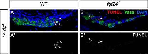
Gonads of wild-type and fgf24 mutant animals have low levels of TUNEL incorporation at 14 dpf. (A-B') TUNEL incorporation and Vasa staining of 14 dpf gonads. Both wild-type (WT; n = 5; A, A') and fgf24 mutant (n = 5; B, B') gonads show similarly low levels of TUNEL staining (red, arrowheads). A-B' are sagittal optical sections with anterior to the left. Germ cells are labeled with Vasa (green), nuclei are labeled with DAPI (blue). Scale bars = 20 μm. |
|
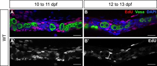
Larval germ cells do not incorporate EdU. (A-B') Single plane confocal micrographs of whole-mount wild-type larval gonads showing EdU incorporation (red). Larvae were allowed to swim freely in 200 μM EdU + 0.1%DMSO from 10 to 11 dpf (A, A') or 12 to 13 dpf (B, B'), euthanized, fixed, and processed for detection of EdU. Many SGCs are EdU-positive at both timepoints, while germ cells are consistently EdU-negative. Germ cells are labeled with Vasa (green) and nuclei are labeled with DAPI (blue). A' and B' show the EdU channel only, in grey. A,-B' are sagittal optical sections with anterior to the left. Scale bars = 20 μm. |
|
Basal laminae are absent from 8 dpf wild-type gonads. (A, A') Single plane confocal micrographs of whole-mount larval gonads immunostained for Laminin (red) and Vasa (green). Laminin is undetectable in either merged (A) or Laminin-only channel (A'), suggesting that basal laminae have not formed. A, A' are sagittal optical sections with anterior to the left. Nuclei are labeled with DAPI (blue). Scale bars = 20 μm. |
|
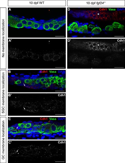
Wild-type and mutant gonads have low levels of membrane-associated Cdh1/E-cadherin at 10 dpf. (A-D') Single plane confocal micrographs of whole-mount larval gonads immunostained for Cdh1/E-Cadherin. In most 10 dpf wild-type (WT; A, A'; 10/15) and fgf24 mutant (D, D'; 10/10) animals, Cdh1 (red) does not localize to cell membranes of gonadal cells. In some cases, wild-type animals have low expression of Cdh1 at the membranes of SGCs (B, B'; 3/15) or germ cells (C, C'; 2/15). A-D' are sagittal optical sections with anterior to the left. Germ cells are labeled with Vasa (green), nuclei are labeled with DAPI (blue). (A', B', C', D) Cdh1 channel only, in grey. Arrow = membrane localization of Cdh1 in gonadal cells; Asterisk = membrane localization of Cdh1 in a nearby, non-gonadal tissue. Scale bars = 20 μm. |
|
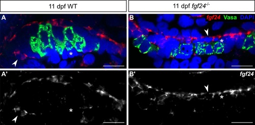
fgf24 expressing and non-expressing somatic cells are present in the gonads of fgf24 mutants. (A-B') Single plane confocal micrographs of whole-mount larval gonads after fluorescent in situ hybridization. fgf24 mRNA (red) can be detected in some, but not all, SGCs of both wild-type (WT; A, A') and fgf24 mutant (B, B') animals at 11 dpf. A-B' are sagittal optical sections with anterior to the left. Germ cells are labeled with Vasa (green), nuclei are labeled with DAPI (blue). Arrowhead = fgf24-positive SGC; Asterisk = fgf24-negative SGC. Scale bars = 20 μm. |

ZFIN is incorporating published figure images and captions as part of an ongoing project. Figures from some publications have not yet been curated, or are not available for display because of copyright restrictions. PHENOTYPE:
|

Unillustrated author statements PHENOTYPE:
|


