
Fig. 7
- ID
- ZDB-FIG-171018-16
- Publication
- Leerberg et al., 2017 - Fibroblast growth factor signaling is required for early somatic gonad development in zebrafish
- Other Figures
- All Figure Page
- Back to All Figure Page
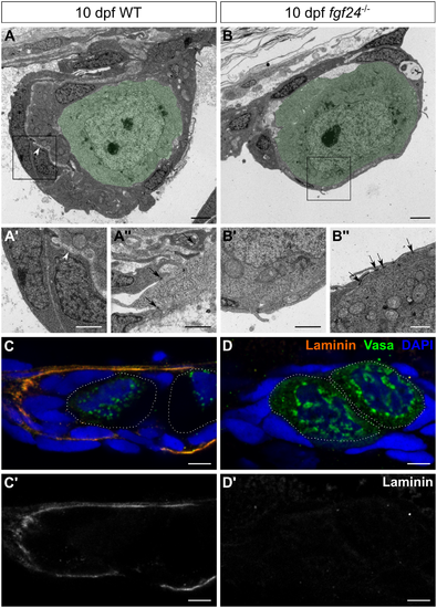
fgf24 mutant gonads lack wild-type organization. (A-B'') Transmission electron micrographs of 10 dpf gonads, transverse sections. In both wild-type (WT) and fgf24 mutant gonads, germ cells (pseudocolored green) are enclosed by SGCs (A, B). However, the somatic gonad component of the wild-type gonad is thicker than in the fgf24 mutant gonad, and is split into two layers by an electron-lucent space (arrowheads in A and A', which is a higher magnification of the box in A). The somatic gonad component is single layered, as shown at higher magnification in B' (corresponds to the box in B). Somatic gonad cells extend processes and make cell-cell contacts (arrows) in both wild-type and fgf24 mutant gonads (A'', B'', respectively). (C-D') Antibody staining against Laminin (orange) and Vasa (green) of 10 dpf gonads; individual germ cells are outlined with dotted white lines. Laminin is deposited between nuclei of SGCs in wild-type (C, C') but not fgf24 mutant (D, D') gonads. C-D' are sagittal optical sections with anterior to the left. Nuclei are labeled with DAPI (blue). Scale bars = 2 μm in A, B; 1 μm in A', A'', B', B''; 5 μm in C-D'. |
| Gene: | |
|---|---|
| Antibodies: | |
| Fish: | |
| Anatomical Terms: | |
| Stage: | Days 7-13 |
| Fish: | |
|---|---|
| Observed In: | |
| Stage: | Days 7-13 |