
Fig. 8
- ID
- ZDB-FIG-171018-17
- Publication
- Leerberg et al., 2017 - Fibroblast growth factor signaling is required for early somatic gonad development in zebrafish
- Other Figures
- All Figure Page
- Back to All Figure Page
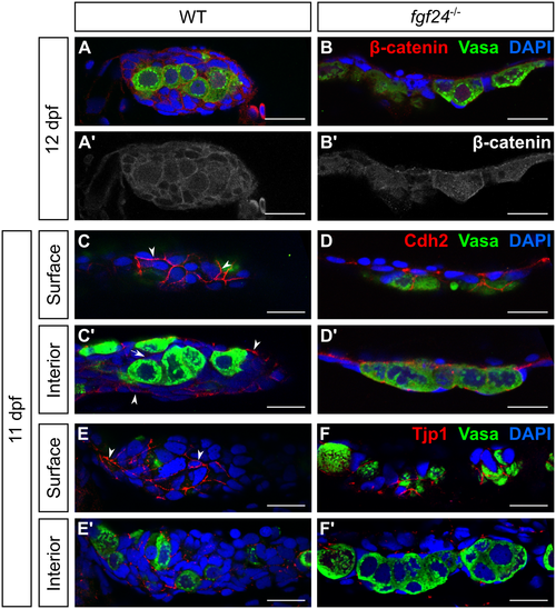
Somatic cells of wild-type and fgf24 mutant gonads express epithelial-like cell adhesion molecules. (A-F') Single plane confocal micrographs of whole-mount larval gonads immunostained for cell adhesion molecules (red). (A-B') β-catenin localizes to the membranes of both SGCs and germ cells in wild-type (WT) gonads (A, A'). While β-catenin still localizes to the membranes of both cell types in fgf24 mutants, it appears reduced (B, B'). (C-D') Cdh2 localizes strongly to the outer layer of SGCs in wild-type and fgf24 mutant gonads, visible on the surface of the gonad (C, D) and in the external-most layer of SGCs of the interior view (C', D'). (E-F') Similar to Cdh2, Tjp1 localizes to the outer layer of SGCs in wild-type (E, E') and fgf24 mutant (F, F') gonads. A-F' are sagittal optical sections with anterior to the left. Germ cells are labeled with Vasa (green), nuclei are labeled with DAPI (blue). Scale bars = 20 μm. |
| Gene: | |
|---|---|
| Antibodies: | |
| Fish: | |
| Anatomical Terms: | |
| Stage: | Days 7-13 |
| Fish: | |
|---|---|
| Observed In: | |
| Stage: | Days 7-13 |