
Fig. S4
- ID
- ZDB-IMAGE-171018-21
- Publication
- Leerberg et al., 2017 - Fibroblast growth factor signaling is required for early somatic gonad development in zebrafish
- All Figures
- Figures for Leerberg et al., 2017
|
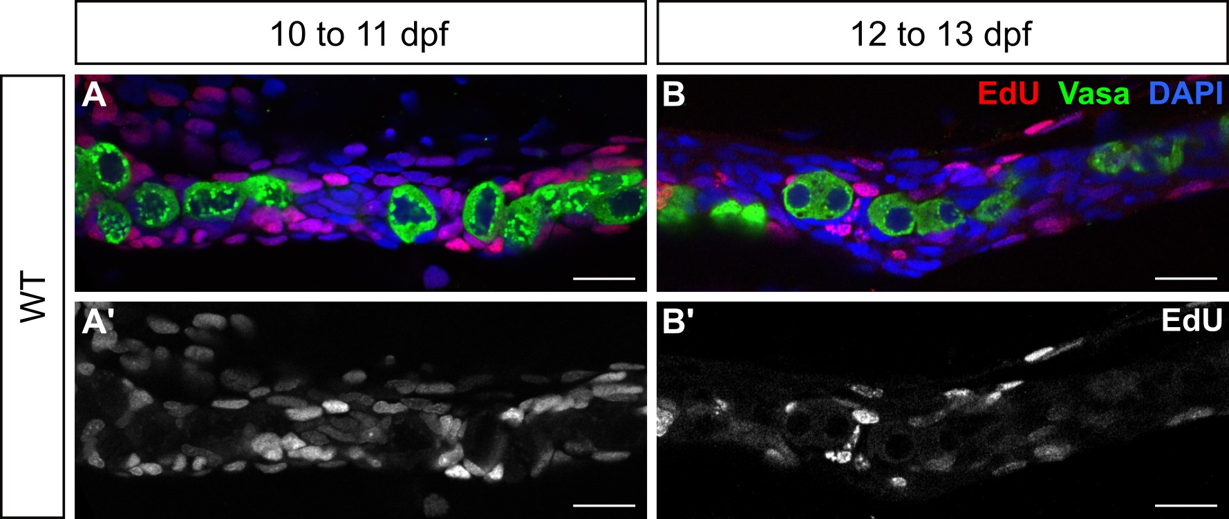
Fig. S4
Larval germ cells do not incorporate EdU.
(A-B') Single plane confocal micrographs of whole-mount wild-type larval gonads showing EdU incorporation (red). Larvae were allowed to swim freely in 200 μM EdU + 0.1%DMSO from 10 to 11 dpf (A, A') or 12 to 13 dpf (B, B'), euthanized, fixed, and processed for detection of EdU. Many SGCs are EdU-positive at both timepoints, while germ cells are consistently EdU-negative. Germ cells are labeled with Vasa (green) and nuclei are labeled with DAPI (blue). A' and B' show the EdU channel only, in grey. A,-B' are sagittal optical sections with anterior to the left. Scale bars = 20 μm.