
Fig. 6
- ID
- ZDB-IMAGE-171018-15
- Publication
- Leerberg et al., 2017 - Fibroblast growth factor signaling is required for early somatic gonad development in zebrafish
- All Figures
- Figures for Leerberg et al., 2017
|
Fig. 6
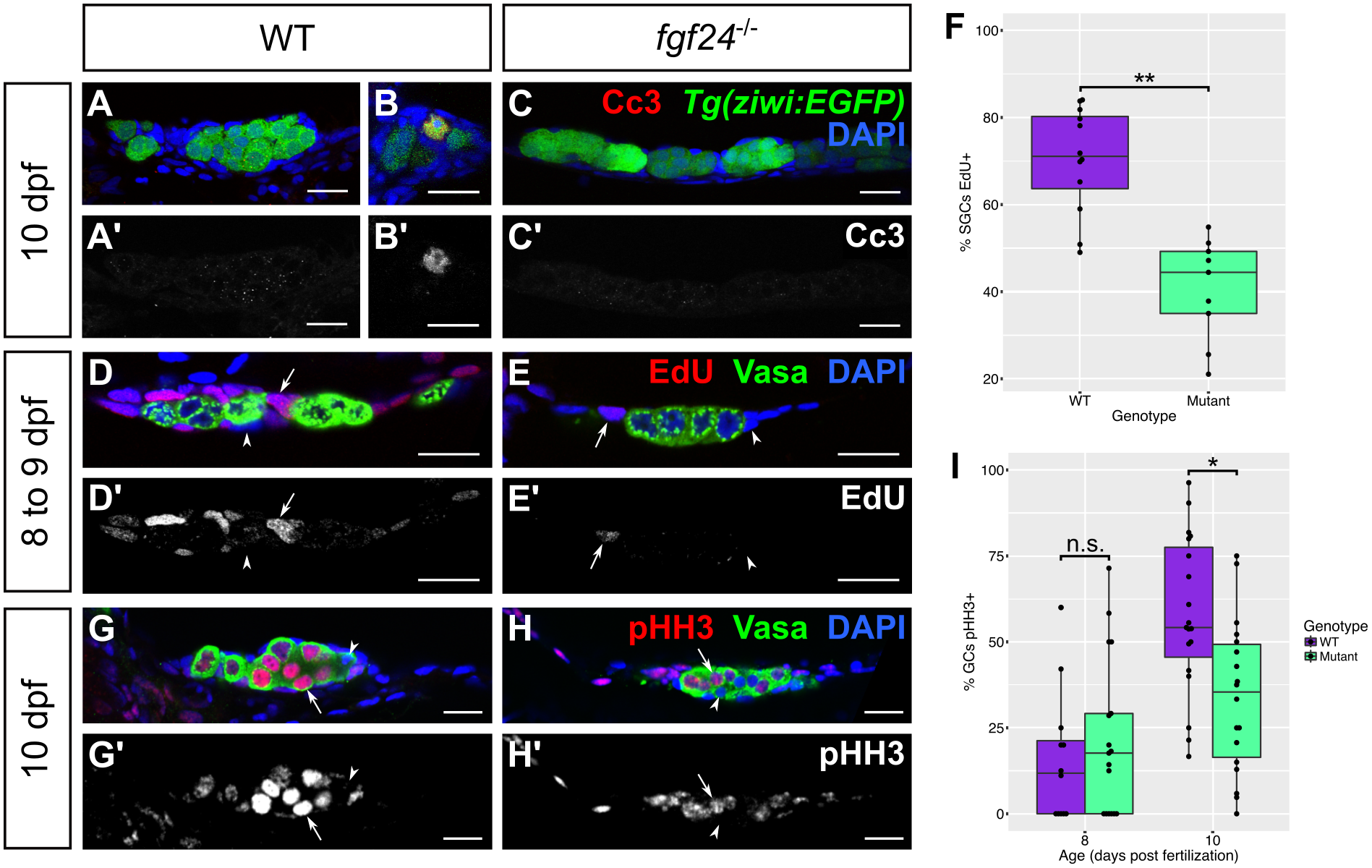
fgf24 mutant somatic gonad cells and germ cells have low proliferation.
(A-C') 10 dpf wild-type (WT; n = 7; A, A') and fgf24 mutant (n = 7; C, C') gonads exhibit rare Cc3 staining (red). Wild-type and fgf24 mutant animals have similar numbers of Cc3(+) germ cells (mean ± SEM = 0.14 ± 0.14, 0, respectively; P = 0.36) and Cc3(+) SGCs (mean ± SEM = 0.57 ± 0.3, 0.43 ± 0.2, respectively; P = 0.7). (B, B') In a 10 dpf wild-type gonad, one Tg(ziwi:EGFP) positive germ cell (green) stains positive for Cc3 (red). (D-F) EdU incorporation from 8 to 9 dpf. Fish exposed to 200 μM EdU between 8 and 9 dpf were euthanized and stained for Vasa (green) and EdU (red). A lower percentage of mutant somatic gonad cells have incorporated EdU compared to wild-type (mean = 41% and 70%, respectively; P < .001; arrow = EdU(+) SGC, arrowhead = EdU(-) SGC). (G-I) Antibody staining against phospho-Histone H3 (pHH3, red) and Vasa (green). At 8 dpf, germ cells of fgf24 mutants are equally likely to be pHH3(+) as those of wild-type animals (mean = 22% and 16%, respectively; P > 0.05), however by 10 dpf, mutants have a significantly lower proportion of pHH3(+) germ cells compared to wild-type (mean = 34% and 58%, respectively; P < .01; arrow = EdU(+) germ cell, arrowhead = EdU(-) germ cell). Nuclei are labeled with DAPI (blue). A-E', G-H' are sagittal optical sections with anterior to the left. (F, I) Box and whisker plots depicting the percentage of proliferative cells in a gonad (purple = wild-type, green = fgf24 mutant). Each dot represents the percentage of EdU(+) SGCs (F) or pHH3(+) GCs (I) in one gonad. (Unpaired two-tailed t-test; * = P < .01; ** = P < .001; n.s. = no significance). Scale bars = 20 μm.