Image
Figure Caption
Fig. S5
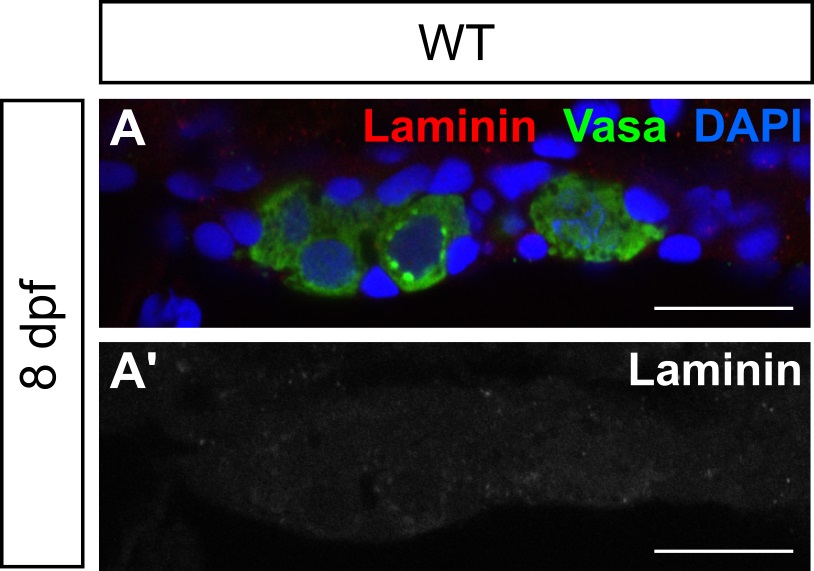
Basal laminae are absent from 8 dpf wild-type gonads.
(A, A') Single plane confocal micrographs of whole-mount larval gonads immunostained for Laminin (red) and Vasa (green). Laminin is undetectable in either merged (A) or Laminin-only channel (A'), suggesting that basal laminae have not formed. A, A' are sagittal optical sections with anterior to the left. Nuclei are labeled with DAPI (blue). Scale bars = 20 μm.
Acknowledgments
This image is the copyrighted work of the attributed author or publisher, and
ZFIN has permission only to display this image to its users.
Additional permissions should be obtained from the applicable author or publisher of the image.
Full text @ PLoS Genet.

