- Title
-
Drl.3 governs primitive hematopoiesis in zebrafish
- Authors
- Pimtong, W., Datta, M., Ulrich, A.M., Rhodes, J.
- Source
- Full text @ Sci. Rep.
|
Drl gene family members are expressed throughout embryonic development and in multiple hematopoietic lineages. (a) RT-PCR analysis of the drl family members and β−actin from pooled embryos at the indicated ages. (b) Semi-quantitative (q) RT-PCR of drl genes and β-actin from pooled 14 somite stage embryos (left panel). Arrows indicate the bands that were used for quantitation, which is shown in the right panel. Compared to drl, *P = 0.0150, **P = 0.0052, EXPRESSION / LABELING:
|
|
Knockdown of the drl gene family causes developmental defects. (a) (Left panel) Diagram comparing exon homology and showing the location of morpholino-targeted sequences. The percent homology for each exon compared to drl is indicated. (Right panel) Alignment of morpholino target sequence (in red) compared to corresponding sequence in each drl family member. DNA base pairs are color-coded. (b) (Upper panel) Individual embryo RT-PCR analysis of drl family members in control and morpholino-injected embryos as indicated. MM indicates a 5 base pair mismatch control morpholino. UI = uninjected. (Lower panels) Quantitation of drl gene family transcripts from RT-PCR analysis. (Left) Control (MO2MM) versus MO3 samples. (Right) Control (L3MM) compared to L3MO samples. Samples were normalized to β-actin. Rel. = Relative. Bars show mean ± S.D. from triplicates; *P = 0.0382, **P = 0.0088, ***P < 0.0001 and EXPRESSION / LABELING:
PHENOTYPE:
|
|
Drl.3 is essential for erythroid development. (a) Control- (mismatch, L3MM) and drl.3 morpholino- (L3MO) injected embryos at 24 hpf showing live Tg(fli1a:EGFP) marking of the vasculature and gata1a and hbae1 WISH, as indicated. (b) Embryos at 35 hpf showing live Tg(fli1a:EGFP) pattern, with a magnified view of the PBI region, and o-dianisidine stained embryos with an enlargement of the anterior region. (c) RT-PCR analysis of individual Tg(gata1:DsRED) control and L3MO-injected embryos. Drl.3 morphants were sorted for normal or decreased (estimated ≤ 60% of normal) numbers of circulating erythrocytes at 48 hpf. Full-length gel images for these cropped panels are provided in Supplemental Figure 8. (d) Quantitation of the levels of drl.3 expression normalized to β-actin from RT-PCR analysis in (c). **P = 0.0019 and **P = 0.0015 (Student's t-test). (e) WISH of gata1a and slc4a1a in 48 hpf control and drl.3 morphants. (f) O-dianisidine stained control and drl.3 morphants at 4 dpf; lateral views (left panels) and ventral views of the anterior region (right panels). (a–b, e–f) The number of the embryos with the representative phenotype per total number of embryos is shown; lateral views with head to the left, dorsal upward. |
|
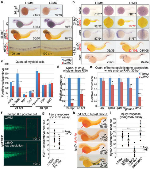
Knockdown of drl.3 transiently decreases myeloid cells without altering the emergence of primitive progenitor and definitive stem cells. (a) WISH of scl at 24 and 48 hpf, and runx1 (dark blue)/slc4a1a (red) at 48 hpf in L3MM- and L3MO-injected embryos, as labeled. Embryos shown as lateral views. (b) WISH of spi1b, l-plastin and mpx at 24 hpf and l-plastin and mpx 48 hpf. Dorsal, anterior (24 hpf only, right panels) and lateral views are shown. (c) Quantitation of the number of the WISH spi1b+, l-plastin+ and mpx+ cells in the anterior of the embryo at 24 hpf and total body l-plastin+ and mpx+ cells at 48 hpf in L3MM (blue) and L3MO-injected (red) embryos (N = 8 for each column except for mpx at 24 hpf where N = 15, bars show mean ± S.E.). **P = 0.0044, ***P < 0.0001 and *P = 0.0156 (Student's t-test). (d) Quantitative real-time PCR analysis of drl.3 in whole embryo RNA samples from 24 and 48 hpf drl.3 morphants (red) and controls (L3MM, blue, set to 1, arbitrary units). **P = 0.0042 and ***P < 0.0001 (Student's t-test). (e) Quantitative real time PCR analysis of scl, spi1b, gata1a, gata1b, and drl.3 in whole embryo RNA samples from pools of 30 hpf drl.3 morphants (red) compared to control-injected embryos (blue, set to 1, arbitrary units). *P = 0.0114, **P = 0.0083 and ***P = 0.0005 (Student's t-test). (d–e) Bars show mean ± S.D., from three independent experiments. Expression was normalized to gapdh. (f) Tail region of spi1:GFP embryos at 54 hpf, 6 hours after tail transection. Selected L3MO embryos had decreased circulating cells; controls were randomly selected, and had normal circulation. Red boxes = tail cut region. (g) Number of spi1:GFP+ cells in tail cut region in control or drl.3 morphants. P = 0.007 (Student's t-test). (h) WISH of lysozymeC/lysC (blue) and slc4a1a (red) at 54 hpf, 6 hours after tails were cut. (i) Number of lysC+ cells in an equal sized region surrounding the tail in the indicated embryos at 54 hpf, 6 hours after tail cuts were performed. P = 6.77E-5, Student's t-test. |
|
L3MO-induced defects are due to knockdown of drl.3 activity. (a) WISH of gata1a at 24 hpf, l-plastin at 25 hpf, and slc4a1a at 48 hpf in L3MM- and L3MO-injected embryos and embryos co-injected with L3MO and drl.3 mRNA. Embryos shown as lateral views, head left. WISH for l-plastin also shows dorsal, anterior views. (b) Percent of L3MM- and L3MO-injected and L3MO/drl.3 mRNA co-injected embryos with normal (wt) or decreased (*; estimated ≤ 60% of normal) numbers of gata1a or slc4a1a-expressing cells. The numbers of embryos are indicated in the columns. ***P < 0.0001 (Fisher's exact test). (c) Percent of embryos with normal (wt) or decreased (*) numbers of l-plastin-expressing cells, as labeled. The numbers of embryos are indicated in the columns. *P = 0.0445 (Fisher's exact test). |
|
Loss of gata1a and spi1b affect drl gene family expression. (a) WISH of drl family members in gata1a and spi1b morphants (MO) compared to uninjected embryos at 24 hpf. From top to bottom: drl, drl.1, drl.2, drl.3, l-plastin (dark blue)/slc4a1a (red) and spi1b. Lateral views, head to the left (left panels); anterior, dorsal views (right panels). The number of the embryos with the representative phenotype out of the total number of embryos is indicated. Arrows in the panels showing dorsal views indicate an increase or decrease in the numbers of WISH+ cells. (b) Quantitation of the number of drl gene family-expressing cells in the anterior hematopoietic region of control (UI), gata1a morphants and spi1b morphants at 24 hpf. N = 10 for each column. Bar shows mean ± S.D. *P = 0.0013, **P = 0.0002, |
|
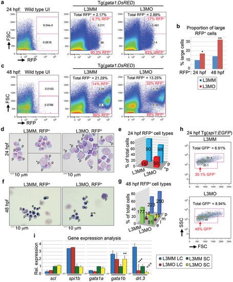
(a) FACS plots of 24 hpf cells from wild-type uninjected (UI) and Tg(gata1:DsRED) embryos injected with L3MM or L3MO as indicated. (b) Quantitation of the percent of large cells RFP+ populations from FACS analysis of L3MM- (blue) and L3MO-injected (red) Tg(gata1:DsRED) embryos, as indicated. *P = 0.0107; **P = 0.0021 (Chi-Squared test). (c) FACS plots of 48 hpf cells from wild-type uninjected (UI) and Tg(gata1:DsRED) embryos injected with L3MM and L3MO as indicated. (a, c) The percent of RFP+ cells out of total cells and the percent of large (FSC high) and smaller sized cell (FSC low) populations are indicated. (d) May-Grunwald-Giemsa (MGG) staining of purified Tg(gata1:DsRED) cells from the indicated 24 hpf morphants. *p = less differentiated progenitor cell; p = progenitor cell, more differentiated. (e) Cell type distribution in purified Tg(gata1:DsRED) cells at 24 hpf based on MGG staining. L3MM versus L3MO (*p and p cells), P = 0.0004 (Fisher's exact test). (f–g) MGG staining of purified 48 hpf Tg(gata1:DsRED) cells (f) and quantitation of MGG cell type distribution (g). p = progenitor cells; e = erythroid cells (≤8 μm); m = myeloid; l = lymphoid. L3MM versus L3MO (e and p cells), P < 0.0001 (Fisher's exact test). The yellow-blue color balance of merged L3MM and L3MO images was slightly adjusted in Photoshop. (e, g) The cell count for each cell type is indicated in the appropriate column. (h) FACS plots of cells from 24 hpf Tg(spi1:EGFP) L3MM- (top) and L3MO-(bottom) injected embryos. An arrow indicates the less mature population (P = 0.0069, Chi-squared test). Supplementary Figure S6a–b shows gating for GFP+ cells. (i) Real-time PCR of scl, spi1b, gata1a, gata1b, and drl.3 in purified large and small size populations of gata1:RFP+ cells from 24 hpf L3MM- and L3MO-injected embryos. The relative expression in small L3MM cells was set to 1, arbitrary units. Expression was normalized to gapdh. Bars show mean ± S.D., triplicate experiments. **P = 0.0083, ***P = 0.0004, and ***P = 0.0009 (Student's t-test). |
|
Drl.3 is expressed in definitive and adult hematopoietic cells. (a) Transverse sections of 28 hpf embryos showing runx1 (left) and drl.3 (right) WISH-positive cells (arrows). Cells in the ventral wall of the aorta are indicated by black arrows. (b-c) WISH analysis of runx1 (blue) and drl.3 (red) in the AGM region of 36 hpf embryos. Runx1 was visualized with NBCIP/NBT (purple), drl.3 by Fast Red alkaline phosphatase substrate. Fast Red precipitate was used to fluorescently visualize drl.3 transcripts. Arrows indicate coexpressing cells. (b) Lateral whole mount views and (c) 8 micron transverse sections showing brightfield (BF, top), fluorescent (IF, middle) and BF/IF overlaid (Merge, bottom) images. (d) Ori family member expression in gata1:GFP+ cells (small sized) purified from adult kidney marrow. (e-f) FAGS analysis of mpx:GFP+ (e) and gata1:RFP+ (f) cells from adult whole kidney marrow (KM). FSC-SSC plots are shown for the fluorescent populations. (g) Real time PCR quantification of scl, gata1a, spi1b and drl.3 expression in gata1:RFP+ large cells (LC) and small cells (SC) from adult KM. ***P = 0.0003, **P = 0.0097. (h) Real time PCR quantification of gata1 and drl.3 expression in gata1:RFP+ and mpx:GFP+ populations as indicated. *P= 0.0425, ***P= 3.78E-05, *P= 0.0115, ^P= 0.0221. Significance was determined using the Student's t-test. |
|
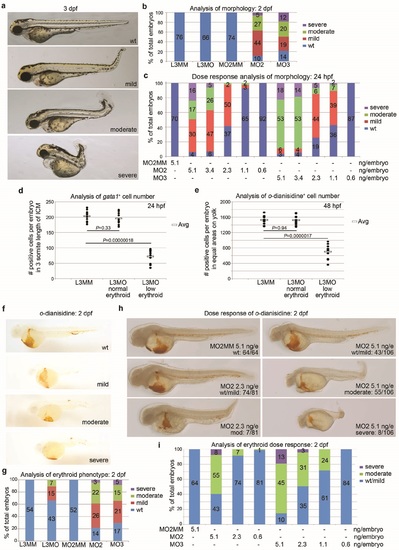
Phenotype analysis of drl family morphants. (a) Brightfield microscopy of embryos at 3 dpf with morphology defined as wild type (wt), mild, moderate, and severe as listed. Representative embryos are control (wt) or M02-injected morphants. (b) Percent of morpholino-injected embryos at 2 dpf that display developmental defects. Phenotypes defined in (a). Control mismatch morpholinos = L3MM and M02MM. The number of embryos is shown in the columns. (c) Embryos injected at the indicated dose were scored for severe, moderate, mild or normal body axis length/amount of tissue as outlined in panel a. Percent of total embryos is charted, with the embryo number shown in the column. (d) Quantitation of 24 hpf gata1-expressing cells in a 3-somite span of ICM (somites 10-13, counting from anterior) in morphants appearing to have normal (L3MM, L3MO normal) or decreased (L3MO decreased) cell numbers as labeled. (e) Quantitation of 48 hpf o-dianisidine stained cells in an equal area and region on the (left side) yolk in morphants appearing to have normal (L3MM, L3MO normal) or decreased (L3MO decreased) cell numbers as labeled. All embryos scored displayed blood pooling on the left side of the yolk. Scatter charts show cell numbers in individual embryos (closed circles); 1 O embryos per group. Average cell number per embryo is indicated (open box). Significance was determined by 2-tailed paired Student's t-test. (f) 0-dianisidine staining of control (wt) and zebrafish M02 morphants. (g) The quantitation of embryos showing wild type, mild, moderate or severe decreases in mature erythroid cells. Phenotypes defined in (f). (h) Representative images of 48 hpf o-dianisidine stained embryos injected with the indicated doses of M02MM and M02. Phenotype scoring is indicated: Wild-type=wt; moderate and severe. Note that the severity of the morphological defects correlate with the severity of the erythroid defect. (i) Quantitation of the embryos with the o-dianisidine phenotypes outlined in (h) injected with the indicated doses of morpholino. |
|
Enforced expression of drl is not sufficient to rescue drl.3 morphants. (a) WISH of drl, drl.1, drl.2, and drl.3 in 24 hpf L3MM- and L3MO-injected embryos. Lateral views, head to the left (right) and dorsal anterior views (left). Number of embryos with representative phenotypes per total embryos analyzed is indicated. (b) Percent of control (L3MM) or drl.3 morphants (L3MO) ± drl mRNA that display normal (wt) or low (estimated s 60% of normal) numbers of o-dianisidine+ cells. Two amounts of drl mRNA were tested: 3 ng/embryo and 6 ng/embryo. Co-injection of L3MO + 3 ng of drl mRNA per embryo resulted in 32% of embryos showing decreased numbers of o-dianisidine+ cells versus 34% of drl.3 morphants. Similarly, 6 ng drl mRNA + L3MO resulted in 32% of the embryos displaying decreased erythrocytes compared to 29% of the L3MO-injected morphants. P values determined by Fisher's exact test. |
|
Knockdown of drl.3 does not alter cell survival or proliferation during embryonic hematopoiesis. (a) Acridine orange staining of L3MMand L3MO-injected embryos at 24 hpf. {b) lmmunofluorescent detection of cleaved Caspase-3 in L3MMand L3MO-injected embryos at 24 hpf. (a-b) Numbers of embryos with representative phenotypes per total embryos analyzed are indicated. (c) Confocal analysis of phospho-Histone H3 (pH3, green) immunodetection and DAPI (blue) staining in L3MM- and L3MO-injected embryos at 22 hpf. Dashed line outlines the ICM. The mitotic index of ICM cells is shown in the right panel. Bars are mean ± S.D. from 8 embryos per condition. |


 , xP = 0.0360,
 , *P = 0.0142, **P = 0.0029 and ***P < 0.001 (Student's t-test). (c) Quantitative real-time PCR analysis of drl.3 in whole embryos at the indicated ages. The relative expression of drl.3 was normalized to the expression of gapdh. (d–e) WISH of drl.2 (d) and drl.3 (e) during embryonic development. Ages of embryos are indicated. Embryos at 2-cell and 30% epiboly stages are shown in a lateral view, animal pole at the top. 50% epiboly embryos are shown in an animal pole view, dorsal to the right. Embryos at 75% and 90% epiboly are shown as lateral views, dorsal to the right. Staged embryos at 10 somites, 18 somites, 24 and 48 hpf are shown as lateral views, anterior to the left. White arrows indicate ALM; asterisks indicate ICM; black arrows indicate PLM; arrowheads indicate AGM cells. Insets show magnified AGM region of corresponding embryo. (f) RT-PCR analysis of the drl family members and β−actin from purified populations of hematopoietic cells. The age of the embryos and the transgenic lines from which cells were purified are indicated. (g) Quantitative real-time PCR analysis of drl.3 in 24 hpf sorted hematopoietic cells. The relative expression of drl.3 was normalized to the expression of gapdh. ***P < 0.0001 (Student's t-test). (b–c, g) Bars show mean ± S.D. (a–b, f) Full-length gel images are provided in Supplemental Figure 8.
 (Student's t-test). (c) Detection of Drl.3 protein and Tubulin in 24 and 48 hpf lysates extracted from uninjected (UI), L3MO- or MO2-injected embryos, 293T cells, and 293T cells expressing Drl.3. (d) Quantitation of Drl.3 from Western blot analysis (from c, normalized to Tubulin). Norm. rel. = normalized relative; AU = arbitrary units. (e) Bright field microscopy of uninjected, control morpholino-injected embryos (MO2MM and L3MM) and morpholino injected-embryos at 18 somites. Lateral views, head to the left, dorsal upward. (f) WISH of scl (dark blue), krox20 (red, rhombomeres 3 and 5), and myoD (red, somites) in 9–10 somite stage embryos. Dorsal views of flat mounted embryos, anterior to left. (e–f) The number of the embryos with the representative phenotype per total number of embryos is indicated for each panel. Full-length gel images and western exposures for the cropped panels (b–c) are shown in Supplemental Figure 8.


 and ***P ≤ 0.0001 (Student's t-test). (c) WISH of drl family members in the indicated embryos at 48 hpf. Embryos shown as lateral views. Horizontal arrows indicate the region where cells in circulation can be visualized. Downward facing arrows indicate decreased WISH+ cell numbers. (d) Percent of L3MM-injected, L3MO-injected, and L3MO/gata1a mRNA co-injected embryos that have normal (blue, wt) or low numbers of erythroid cells (red, *estimated ≤ 60% of normal) based on o-dianisidine staining at 48 hpf. (e) Quantitative analysis of uninjected, gata1a MO-injected, and gata1a MO/drl.3 mRNA co-injected embryos that have normal (blue, wt) or low numbers of erythroid cells (red, *) based on o-dianisidine staining at 48 hpf. (d–e) Numbers of embryos are indicated in the columns. Statistical significance was analyzed using Fisher's exact test.


