
- Title
-
The mother superior mutation ablates foxd3 activity in neural crest progenitor cells and depletes neural crest derivatives in zebrafish
- Authors
- Montero-Balaguer, M., Lang, M.R., Sachdev, S.W., Knappmeyer, C., Stewart, R.A., De La Guardia, A., Hatzopoulos, A.K., and Knapik, E.W.
- Source
- Full text @ Dev. Dyn.
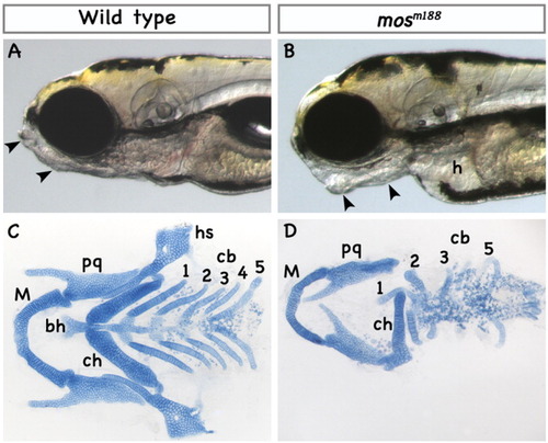
The mother superior (mosm188) mutation affects zebrafish craniofacial development. A,B: Lateral view of wild-type (A) and mosm188 (B) live embryos at 4 dpf. In mosm188 embryos, the Meckel's cartilage is pointing ventrally and the posterior arches are missing (arrowheads point to the anterior and posterior tips of the Meckel's cartilage). The heart (h) develops an edema. C,D: Ventral view of dissected pharyngeal cartilages stained with Alcian blue from wild-type (C) and mosm188 (D) embryos at 4 dpf. Cartilage elements corresponding to second and posterior arches (marked 1-5) are severely reduced. cb, ceratobranchial; bh, basihyal; ch, ceratohyal; h, heart; hs, hyosymplectic; M, Meckel's cartilage; pq, palatoquadrate. PHENOTYPE:
|

The mosm188 mutation affects migratory cranial neural crest. A,B:dlx8 expression was analyzed by in situ hybridization at 36 hpf in the craniofacial primordia of wild-type (A) and mosm188 (B) embryos. In mosm188mutants, expression appears slightly reduced in the first primordium but it is faint in the second (arrow in all panels) and absent in the posterior streams. C,D: Dorsal view of dlx2 expression at 24 hpf shows markedly reduced expression in the first two neural crest streams and almost complete absence in the postotic ones. E,F: Expression of the paraxial mesoderm and head mesenchyme marker wnt5a is substantially reduced at 24 hpf in the second and posterior NC streams in the mosm188 mutants while the first arch expression appears unaffected. G,H: The expression of sox9a, a marker of cranial mesenchymal condensations, is reduced in the first arch and is completely abolished in the second and posterior arches (24 hpf). I-L: The hox gene expression in hindbrain rhombomeres (r) is comparable between wild-type (I, K) and mosm188 (J, L) embryos, but expression of hoxa2 in the hyoid neural crest stream is largely absent, (arrow, J). Expression of hoxa2 and hoxb3 in the postotic stream is considerably reduced (white arrowhead in I,L). |
Pigment cells are differentially affected by the mosm188 mutation. A,B: At 36 hpf, the rostrocaudal distribution (arrow) and number of melanophores are delayed in mosm188 as compared to wild-type embryos. C,D: The number and distribution of melanophores in mosm188 mutants at 3 dpf is comparable to wild types. E,F: The number of trunk iridophores (arrows) is reduced in mosm188 mutant embryos at 3.5 dpf, while the eye iridophores are not affected. Iridophores were photographed under incident light, dorsal views. Arrowhead points to the iridophore precursor patch in the trunk. PHENOTYPE:
|
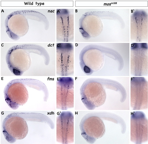
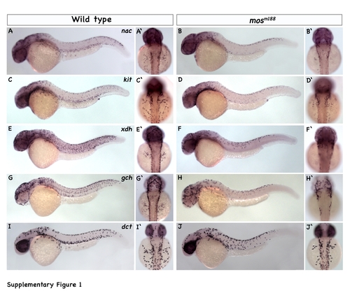
Pigment cell progenitors show deficits in the mosm188 mutants. Expression analysis of pigment specific genes in wild type (A, C, E, G) lateral and (A', C', E', G') dorsal views, and in mosm188 mutants (B, D, F, H) lateral and (B', D', F', H') dorsal views as revealed by whole mount in situ hybridization in 22 hpf embryos. A-B': Expression of mitfa/nac, the early marker of pigment progenitors, is significantly downregulated in the trunk region while the mid-hindbrain boundary domain is unaffected in mosm188 mutants. C-D': Similarly, the melanophores specific dct/trp2 expression is largely absent in mosm188 mutants at this stage. E-F':Fms that specifies melanophore and xanthophore lineages is essentially absent in mosm188mutants as is xanthine dehydrogenase (G-H';), the enzyme in pteridine synthesis pathway that marks lineage-committed xanthoblasts. EXPRESSION / LABELING:
|
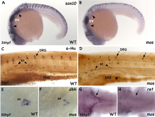
Neural derivatives of NC are affected by the mosm188 mutation. A,B: In wild types (A) at 24 hpf, sox10 is expressed in late specified pigment and neural precursors migrating ventrally from the dorsal aspect of the neural tube, in the otic vesicle (ov), and in the preotic and postotic clusters of glial precursors (arrowheads). In the mosm188 mutant embryos (B), the otic vesicle expression is unchanged, while expression in all other domains is greatly reduced. C,D: Anti-Hu staining in the trunk of 4-dpf embryos. The wild-type pattern of the segmentally organized DRGs (arrows), enteric neurons (ENS), and sympathetic neurons (sn; arrowheads) is shown in C. In the mosm188 mutant (D), there are only a few scattered DRGs (arrows) and sympathetic neurons (arrowheads). An asterisk marks the area devoid of enteric neurons in the digestive tube. E,F: The reduction of the sympathetic neuron precursors in mosm188 mutants is already evident at 50-hpf embryos stained with the dopamine beta hydroxylase (dbh) riboprobe. G,H: The enteric neuron precursors migrate into the gut tube at 36 hpf as marked by ret-positive cells in wild-type embryos (G). The ret expression is greatly reduced in mosm188 mutants (H). DRG, dorsal root ganglia; ENS, enteric nervous system; ov, otic vesicle; sn, sympathetic neurons. |
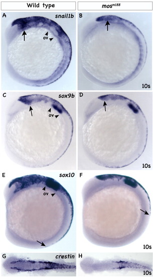
Expression of NC specification genes. A,B: Expression of snail1b in the premigratory NC cells (arrow) of wild-type embryos (A) is localized to the dorso-lateral edge of the neural tube, but it is greatly reduced in mosm188 mutants (B). The expression in the somites is unaltered. The arrowheads in wild-type embryos demarcate the position of the otic vesicle. C,D:Expression of sox9b is greatly reduced in NC progenitors (arrows) in mosm188 mutants (D), while the otic vesicle expression (arrowheads) is unaltered. E,F: Expression of sox10 in premigratory NC cells of wild-type embryos (E) is localized to the dorso-lateral edge of the neural tube (arrow). Sox10 expression is greatly reduced in mosm188 mutants (F), while the otic vesicle expression (arrowheads) is unaltered. G,H: The wild-type pattern (G) of crestin expression in the cranial region is absent in mosm188 mutants (H), but it is only reduced in the trunk. Lateral views in A-F and dorsal flat mounts in G and H, anterior to the left. EXPRESSION / LABELING:
|
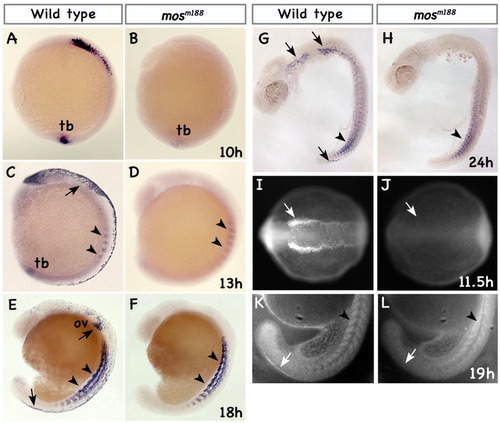
mosm188 mutants lack foxd3 expression in NC precursor cells. Lateral views of wild-type embryos and mosm188 mutants stained with a foxd3 riboprobe (anterior is to the left). A,B:foxd3 expression at the tail bud stage (10 hpf) in wild-type embryos (A) is localized to the future hindbrain cranial neural crest precursors and to the tail bud domain (tb). foxd3 is not expressed in the NC precursors in mosm188 mutants (B) and only faintly in the tail bud domain. C,D: In wild-type embryos (C), cranial and trunk premigratory neural crest cells express foxd3 at the 8-somite stage (13 hpf; arrow points to the posterior edge of the otic placode). Expression is also visible in the somitic precursor cells (arrowheads), and in the tail bud (tb). Expression in mosm188 mutants (D) is restricted to the somites (arrowheads). E,F: The foxd3 message levels in wild types (E) is downregulated in the cranial NC, but it is still maintained in the trunk premigratory NC at the 18-somite stage (arrow, 18 hpf), in the somites (arrowheads), and in the late migrating neural crest precursors around the otic vesicle (ov, arrow). In mosm188 (F), expression is only detected in the somites (arrowheads). G,H: At 24 hpf, decreasing numbers of cells express foxd3 in the most posterior somites of wild-type embryos (Lee et al.,[2006]; arrowhead in G). At this time, foxd3 expression of the NC is confined to the two domains located anterior and posterior to the otic vesicle (arrows) that will contribute to neural derivatives. In the mosm188 mutants (H), the somites continue expressing foxd3 (arrowhead), while there is no expression in the cranial and trunk NC progenitors. I,J: The antibody against zebrafish Foxd3 protein marks the two domains at the neural plate border (arrow) where NC progenitors are located at 11.5 hpf (4-somites) in wild types, while there is no detectable protein in mosm188 mutant embryos at this stage (J). K,L: At 19 hpf, wild-type but not mosm188 mutant neural crest, seen at the most distal part of the tail, express Foxd3 protein, while there is no difference in somitic expression. EXPRESSION / LABELING:
|

Knockdown of the Foxd3 protein phenocopies mosm188 defects. A,B: Dorsal views of 4-somite stage embryos stained by immunohistochemistry using a zebrafish specific anti-Foxd3 antibody. In wild-type embryos (A), Foxd3 protein is localized to two parallel stripes of neural crest precursors at the edge of the neural tube (arrowheads). This pattern is lost in the foxd3 morphants (wild-type embryos injected with a cocktail of two foxd3 morpholinos) corroborating the effective and complete block of foxd3 translation (B). C,D: Lateral views of a live wild type (C) and a foxd3 morphant (D) at 5 dpf. The morphant phenotype closely resembles mosm188 mutant embryos with a gaping jaw and loss of posterior arches (arrows). E,F: Flat mounted Alcian blue stained cartilage preparations of a wild type (E) and a foxd3 morphant (F) show normal Meckel's (M) and palatoquadrate cartilages (pq). The second arch hyosymplectic (hs) in most cases is missing the hyomandibular portion. The ceratohyals (ch) are present, but the posterior pharyngeal arch cartilages are reduced to a few clusters of chondrocytes (arrows). The ceratobranchial (cb) cartilages are malformed. G,H: Expression of snail1b in neural crest precursors (arrows) is greatly reduced in the morphants (H). I,J: This result is concordant with reduced expression of migratory neural crest labeled with dlx2 (arrow points at second neural crest stream). K: Images of live wild-type and morphant embryos under incident light show overall reduced number of fluorescing iridophores in the lateral patch (arrowhead) and in the trunk (arrows) of morphants. EXPRESSION / LABELING:
PHENOTYPE:
|

Chromosomal localization of the mosm188 mutation. A: Positional cloning of the locus disrupted by the mosm188 mutation. The LG6 genomic region is drawn to depict the position of the mutation with respect to microsatellite markers (Z10183, Z15259, Z12094), STSs (BO3194R/F, fc06402), and the foxd3 gene. The distance of each marker from the mosm188 mutation is shown as the number of recombinants in 3,134 meioses. B: The PAC clones D, E, G, and the BAC clone 148H10 contain the foxd3 gene and the mosm188 critical interval. The PAC and BAC clones are aligned to scale and the full sequence of the BAC clone is available from NCBI (Zebrafish Genome Sequencing Project). The four genes within this area and their orientation are marked. C-F: Lateral (C) and dorsolateral (D-F) views of 18-hpf embryos stained with the foxd3 riboprobe. Foxd3 is expressed in wild-type embryos in cranial neural crest (arrow) and somites (arrowheads), while it is expressed only in somites in mosm188 mutants (D). PAC-injected embryos show partial restoration of the foxd3 expression in the cranial region (arrow, E) or unilateral clusters of foxd3-positive cells (arrow, F). G.H: Alcian Blue preparations of 5-dpf pharyngeal skeletal elements in mosm188 (G) and PAC-injected mosm188 embryos (H) show well-differentiated skeletal elements of the 2nd arch (arrow) and posterior arch cartilages. Abbreviations as in Figure 1. PHENOTYPE:
|
Migratory pigment cells recover in the mosm188 mutant embryos. Migratory pigment precursors in wild type (A, C, E, G, I) lateral and (A?, C?, E?, G?, I?) dorsal views, and in mosm188 mutants (B, D, F, H, J) lateral and (B?, D?, F?, H?, J?) dorsal views as revealed by whole mount in situ hybridization of 30 hpf embryos. Although expression patterns in mosm188 mutants lack behind their wild-type siblings, the differences are less pronounced than at 22 hpf in Fig. 4. (A-B?) Expression of mitfa, the early marker of pigment progenitors is present in the most distally and ventrally located cells in wild types and in mos mutants, but in mosm188 expression is reduced in the anteroposterior and dorsoventral extent. (C-D?) The expression of the mitfa target gene kit is only slightly reduced in mosm188 mutants as the migrating cells begin to spread over the yolk (C?, D?). (E-F?) In contrast, expression of the xanthine dehydrogenase gene in lineage-committed xanthoblasts still lacks behind in mosm188 mutants. (G-H?) A similar defect is evident in expression of the GTP-cyclohydrolase, a critical enzyme in the pteridine and melanine synthesis. (I-J?) The dct/trp2 gene that is largely absent in mos at 22 hpf (Fig. 4) is extensively expressed in migratory precursors in the mutant embryos at 30 hpf. EXPRESSION / LABELING:
|
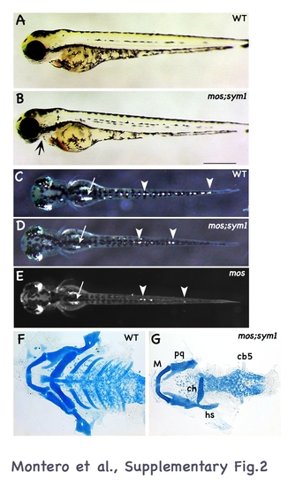
The mosm188 and the foxd3 mutation sym1 do not complement. (A, B) Lateral views of live transheterozygous mosm188;sym1 embryos show loss of posterior arches and a gaping jaw phenotype at 4 dpf (arrow in B). (C, D, E) The auto-fluorescing iridophores are greatly reduced in mosm188;sym1 especially in the trunk (arrowheads) and in the lateral patch (arrow). This phenotype is similar to the mosm188 mutants in E. (F-G) Alcian blue preparations of mosm188;sym1 closely resemble the mosm188 cartilage deficits (in Fig. 1C,D). F-G, flat-mounted preparation of the pharyngeal skeleton at 4 dpf. PHENOTYPE:
|
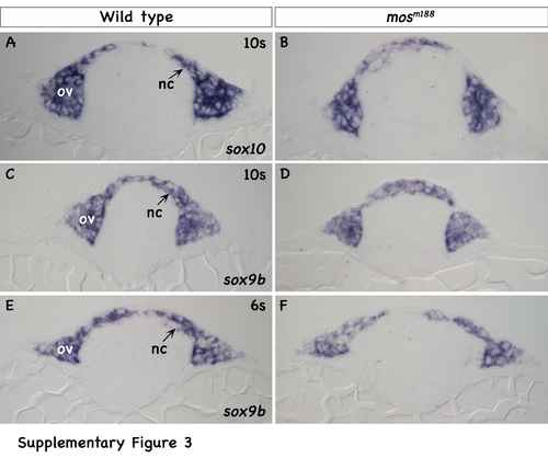
Histological sections reveal comparable populations of NC cells in mosm188 and wild-type siblings. Histological cross-sections of riboprobe labeled embryos at the level of otic vesicle (ov). The populations of sox10 (A,B), and sox9b (C,D) positive neural crest cells at the 10-somite stage at the level of the otic vesicle are similar between wild-type and mutant embryos. The similarity is also evident at an earlier, 6-somite stage (E,F). Embryos were imbedded in JB4 plastic resin and sectioned 5 μm thick. EXPRESSION / LABELING:
|

Unillustrated author statements PHENOTYPE:
|