- Title
-
The Warburg Effect and lactate signaling augment Fgf-MAPK to promote sensory-neural development in the otic vesicle
- Authors
- Kantarci, H., Gou, Y., Riley, B.B.
- Source
- Full text @ Elife
|
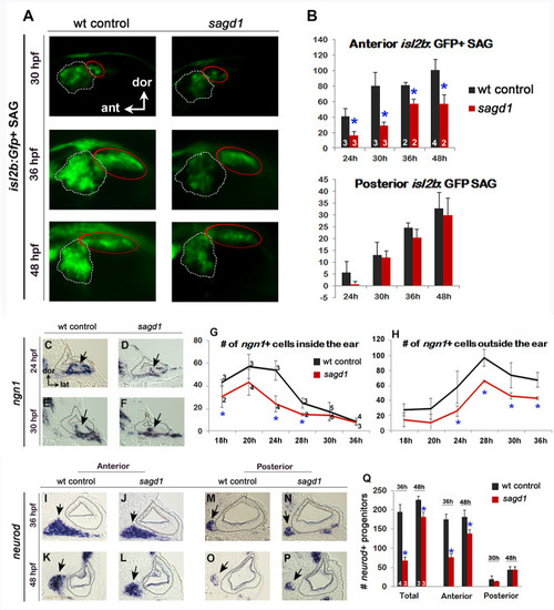
Initial analysis of sagd1.(A) Lateral views of isl2b:Gfp+ SAG neurons in live wild-type (wt) embryos and sagd1mutants at the indicated times. The anterior/vestibular portion of the SAG (outlined in white) is deficient in sagd1 mutants, whereas the posterior SAG (outlined in red) appears normal. (B) Number (mean and s.d.) of isl2b:Gfp+ anterior neurons and posterior neurons in wild-type and sagd1 embryos at the times indicated. Sample sizes are indicated. Asterisks, here and in subsequent figures, indicate significant differences (p<0.05) from wild-type controls. (C–F) Cross-sections through the anterior/vestibular portion of the otic vesicle (outlined) showing expression of ngn1 in wild-type embryos and sagd1 mutants at 24 and 30 hpf. (G, H) Mean and standard deviation of ngn1+ cells in the floor of the otic vesicle (G) and in recently delaminated SAG neuroblasts outside the otic vesicle (H), as counted from serial sections. (I–P) Cross-sections through the anterior/vestibular and posterior/auditory regions of the otic vesicle showing expression of neurod in transit-amplifying SAG neuroblasts at 36 and 48 hpf. (Q) Mean and standard deviation of neurod+ SAG neuroblasts at 30 and 48 hpf counted from serial sections. Sample sizes are indicated (B, G, Q). |
|
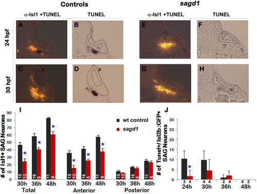
Quantitation of mature Isl1/2+ SAG neurons and TUNEL in sagd1 mutants.(A–H) Cross sections (dorsal up, medial to the right) through the anterior/vestibular portion of the otic vesicle in control embryos (A–D) and sagd1 mutants (E–H) showing anti-Isl1/2 stained SAG neurons (A,C, E, G) and co-staining for TUNEL (B, D, F, H). (I) Number of Isl1/2+ SAG neurons at 30 hpf counted from whole mount preparations. Anterior/vestibular SAG neurons are under-produced in sagd1 mutants, whereas posterior/auditory neurons develop normally. (J) Number of TUNEL+ apoptotic cells at the indicated times. sagd1 mutants show fewer apoptotic cells than normal at 24 and 30 hpf, indicating that the deficiency in vestibular SAG neurons is not due to cell death. Sample sizes are indicated (I, J). |
|
Sensory development and early Fgf signaling are impaired in sagd1 mutants.(A–D”, F–T’) Dorsolateral views of whole mount specimens (anterior to the left) showing expression of the indicated genes in the otic vesicle (outlined) at the indicated times in wild-type embryos and sagd1 mutants. (E) Mean and standard deviation of hair cells in the anterior/utricular and posterior/auditory maculae at 36 and 48 hpf in wild-type embryos (black) and sagd1 mutants (red). Sample sizes are indicated. EXPRESSION / LABELING:
PHENOTYPE:
|
|
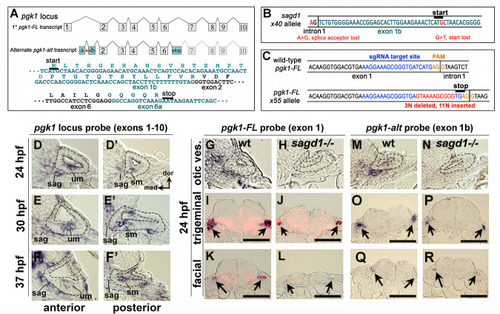
Two independent transcripts associated with the pgk1 locus.(A) Exon-intron structure of the primary full-length pgk1 transcript (pgk1-FL) and alternate transcript (pgk1-alt) arising from an independent transcription start site containing two novel exons (1a and 1b) that splice in-frame to exon 2, and a third novel exon (6a) containing a stop codon. The nucleotide and peptide sequences of exons 1b and 6a are shown. Relative positions of the lesions in sagd1 affecting exon 1b are indicated (red asterisks). (B) Nucleotide sequence near the 5’ end of exon 1b showing the SNPs detected in sagd1 (red font). (C) Nucleotide sequence of pgk1-FL showing the sgRNA target site (blue font) and the altered sequence of the x55 mutant allele (red font), which introduces a premature stop codon. (D–F’) Cross sections through the otic vesicle (outlined) showing staining with ribo-probe for the entire pgk1 locus (covering both pgk1-FL and pgk1-alt) in wild-type embryos. Note elevated expression in the SAG, utricular macula (um), and saccular macula (sm). (G, H, M, N) Cross sections through the otic vesicle (outlined) in wild-type embryos and sagd1 mutants stained with ribo-probe for exon 1 (pgk1-FL alone) (G, H) or exon 1b (pgk1-alt alone) (M, N). Accumulation of both transcripts is dramatically reduced in sagd1 mutants. (I–L, O–R) Cross sections through the hindbrain (dorsal up) showing expression of pgk1-FL (black) plus neurod (red) (I–L) or pgk1-alt alone (O–R). Arrows indicate positions of the trigeminal and facial ganglia. Scale bar, 100 µm. |

ZFIN is incorporating published figure images and captions as part of an ongoing project. Figures from some publications have not yet been curated, or are not available for display because of copyright restrictions. PHENOTYPE:
|
|
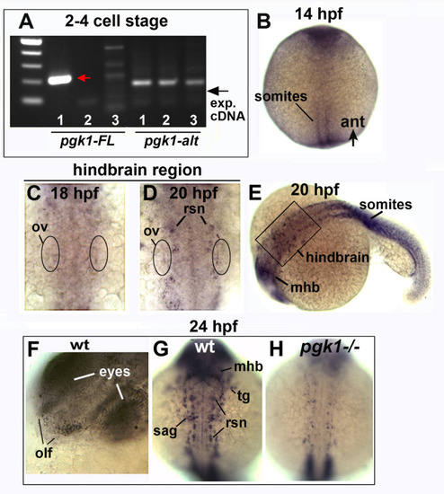
Expression of pgk1-FL during development.(A) RT-PCR for pgk1-FL and pgk1-alt conducted on mRNA harvested at the 2–4 cell stage (lane 1), mRNA samples excluding reverse transcriptase (lane 2), or genomic DNA (lane 3). The expected 211 bp amplicon was obtained for pgk1-FL (red arrow), but not the 163 bp amplicon (exp. cDNA) for pgk1-alt (black arrow). (B–H) Whole mount in situ hybridization for pgk1-FL at the indicated times. Images show dorsal views with anterior to the top (B–D, G,H) or lateral views with anterior to the left (E, F). Expression of pgk1-FL shows the first signs of upregulation in developing somites at 14 hpf (B), whereas diffuse upregulation becomes evident in the hindbrain region beginning at 18 hpf (C) and becomes more intense in developing reticulospinal neurons (rsn) by 20 hpf (D). The position of the otic vesicle (ov) is indicated. The specimen in (D) is depicted at lower magnification in (E) and shows that expression of pgk1-FL also increases in the midbrain-hindbrain border (mhb) and somites. (F–H) Expression of pgk1-FL at 24 hpf in wild-type embryos (F, G) and a pgk1- / - mutant (H). In wild-type embryos local upregulation increases near sites of Fgf expression, including the olfactory epithelium (olf), SAG, midbrain-hindbrain border (mhb), the trigeminal ganglion (tg) and reticulospinal neurons (rsn). Expression is strongly reduced in pgk1- / - mutants. EXPRESSION / LABELING:
PHENOTYPE:
|
|
pgk1-alt misexpression increases pgk1 transcript abundance.(A–N) Expression of pgk1-alt (A–H) and pgk1 (I–N) following heat shock (39°C for one hour) of transgenic and wild-type embryos at 20 hpf. Images show whole embryos (A–F, I–L) or close-ups of the hindbrain (anterior to the top) (G, H, M, N). The position of the otic vesicle (ov) is indicated. Expression of pgk1-alt peaks at 21 hpf in transgenic embryos but decays to normal levels by 24 hpf. In contrast, expression of pgk1 remains elevated in transgenic embryos through 24 hpf. |
|
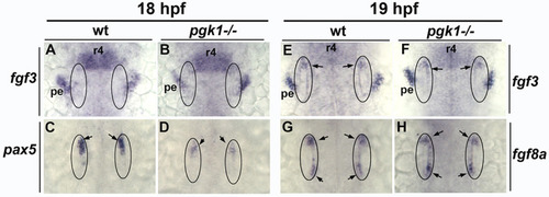
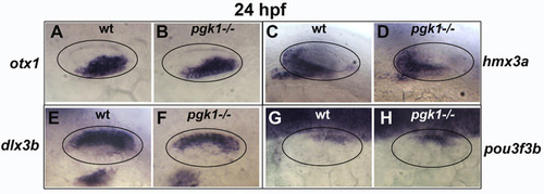
Initial characterization of pgk1- / - mutants and genetic mosaics.(A–F) Cross sections through the anterior/vestibular region of the otic vesicle (outlined) showing expression of the indicated genes in wild-type embryos and pgk1- / - mutants at the indicated times. (G–J) Lateral views showing isl2b-Gfp expression in live embryos at 30 hpf (G, H) and fgf3 at 24 hpf (I, J). The otic vesicle is outlined. (K–P) Cross sections through the otic vesicle (outlined) showing positions of lineage labeled wild-type cells (red dye) transplanted into a wild-type host (K, M, O) or a pgk1- / - mutant host (L, N, P). Sections are co-stained with DAPI (M, N) and etv5b probe (O, P). Note the absence of etv5bexpression in wild-type cells transplanted into the mutant host (P). (Q) Number of mature Isl1+ SAG neurons at 30 hpf in embryos with the indicated genotypes, except for sagd1 x pgk1 intercross expected to contain roughly 25% each of +/+, sagd1/+, pgk1/+ and sagd1/pgk1 embryos. (R) Percent of wild-type donor cells located in the ventral half of the otic vesicle expressing etv5b in wild-type or pgk1- / - hosts. A total of 151 wild-type donor cells were counted in eight otic vesicles of wild-type host embryos, and 247 wild-type donor cells were counted in eight otic vesicles of pgk1- / - host embryos. (S) Number of Isl1+ SAG neurons (total, anterior and posterior) in wild-type and pgk1- / - mutant embryos at 36 hpf (n = 12), 48 hpf (n = 12) and 120 hpf (n = 4). (T) Number of phalloidin-stained hair cells (anterior/utricular and posterior/saccular) in wild-type and pgk1- / - mutant embryos at 36 hpf (n = 16), 48 hpf (n = 16) and 120 hpf (n = 10). Sample sizes are indicated (S, T). EXPRESSION / LABELING:
PHENOTYPE:
|
|
( |
|
( |
|
( |

ZFIN is incorporating published figure images and captions as part of an ongoing project. Figures from some publications have not yet been curated, or are not available for display because of copyright restrictions. PHENOTYPE:
|
|
( |

ZFIN is incorporating published figure images and captions as part of an ongoing project. Figures from some publications have not yet been curated, or are not available for display because of copyright restrictions. PHENOTYPE:
|

ZFIN is incorporating published figure images and captions as part of an ongoing project. Figures from some publications have not yet been curated, or are not available for display because of copyright restrictions. PHENOTYPE:
|
|
Dorsolateral view (anterior to the left) of the otic vesicle (outlined) showing expression of |
|
( EXPRESSION / LABELING:
PHENOTYPE:
|

Unillustrated author statements PHENOTYPE:
|

