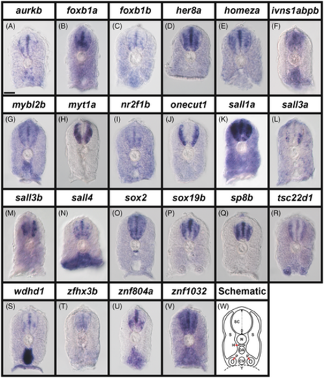
Broad expression of transcription factor and transcriptional regulator genes in zebrafish spinal cord at 24 h. (A–V) Cross-section views of trunk expression of transcriptional regulator gene (A) aurkb, and transcription factor genes (B) foxb1a, (C) foxb1b, (D) her8a, (E) homeza, (F) ivns1abpb, (G) mybl2b, (H) myt1a, (I) nr2f1b, (J) onecut1, (K) sall1a, (L) sall3a, (M) sall3b, (N) sall4, (O) sox2, (P) sox19b, (Q) sp8b, (R) tsc22d1, (S) wdhd1, (T) zfhx3b, (U) znf804a, and (V) znf1032 in WT zebrafish embryos at 24 h. Dorsal, up. A minimum of five embryos were analyzed per gene to determine the representative expression pattern (see Experimental Procedures). As indicated in the schematic cross-section (W), the spinal cord (SC), is located above the notochord (N), which is above the hypochord (H, indicated with red arrow), dorsal aorta (DA), and the cardinal vein (CV). The somites (S) can be seen on both sides of these tissues and the pronephros tubes (P, indicated with red arrows) are ventral, either side of the cardinal vein. Within the spinal cord, the dotted line indicates the midline, the small oval indicates the central canal and the small black triangles indicate the roof plate and floor plate. (N) sall4 in situ hybridization experiments were performed with the molecular crowding reagent Dextran Sulfate (see Experimental Procedures for rationale). All other in situ hybridization experiments in this figure were performed without this reagent. Scale bar: 30 μm.
|
