
- Title
-
Convergence of signaling pathways underlying habenular formation and axonal outgrowth
- Authors
- Roberson, S., Halpern, M.E.
- Source
- Full text @ Development
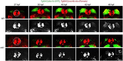
Live imaging of dbx1b+ and cxcr4b+ populations. Live imaging of WT and wls mutant TgBAC(dbx1b:GFP) and TgBAC(cxcr4b:nls-dTomato) double-labeled embryos between 27 and 48 hpf (Movie 1). Black and white images are of dTomato-labeled nuclei alone. dTomato-labeled nuclei are found in the pineal anlage (dashed circle) and, over time, in cells arising within the developing dHb (arrowheads and inset). Scale bar: 30 µm (11 µm for inset). |
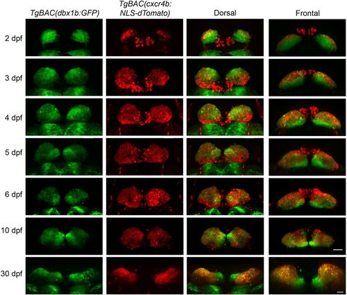
Temporal switch in dbx1b and cxcr4b expression in the developing dHb. Relative positions of dbx1b+ and cxcr4b+ cell populations revealed by TgBAC(dbx1b:GFP) and TgBAC(cxcr4b:nls-dTomato) labeling, respectively, between 2 and 30 dpf. Merged images are shown in dorsal and frontal views. Scale bars: 30 µm. EXPRESSION / LABELING:
|
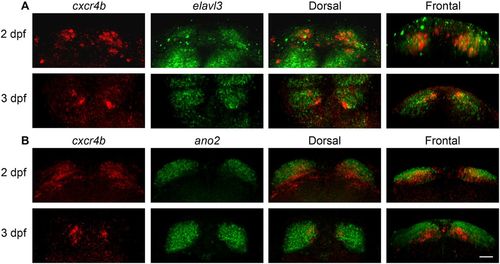
Ventromedial position of cxcr4b-expressing cells relative to dHb neurons. Fluorescence in situ hybridization in 2 and 3 dpf larvae for transcripts of cxcr4b and (A) the pan-neuronal gene elavl3 or (B) ano2, a marker of dHb neurons. The left three columns of images are corresponding dorsal views; the rightmost column shows frontal views. Scale bar: 30 µm. EXPRESSION / LABELING:
|
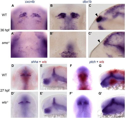
Specification of dbx1b+ dHb progenitors requires Shh signaling upstream of or parallel to Wnt signaling. At 36 hpf, (A,A′) cxcr4b and (B-C′) dbx1b transcripts are found in the developing dHb of (A,B,C) WT embryos, but not in the corresponding diencephalic region of (A′,B′,C′) smo mutants (arrowheads). Expression of (D-E′) shha and (F-G′) ptch1 is comparable in (D,E,F,G) WT embryos and (D′,E′,F′,G′) wls mutants. Sibling WT and homozygous wls mutant embryos are distinguished by the presence or absence of wls transcripts (Kuan et al., 2015). Dorsal (A-B′,D,D′,F,F′) and lateral (C,C′,E,E′,G,G′) views are shown. Scale bars: 30 µm. |
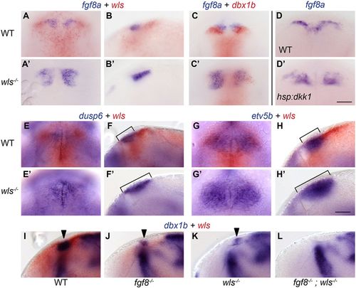
Wnt signaling defines the domain of Fgf signaling in the dorsal diencephalon. (A-C′) At 35 hpf, the domain of fgf8a expression just rostral to the developing dHb is expanded in wls mutants. (D,D′) Inhibition of Wnt signaling at 36 hpf by heat shock activation of Tg(hsp70:dkk1-GFP) also expands fgf8a expression at 48 hpf. (E-H′) The expression domains of (E,F) dusp6 and (G,H) etv5b, two targets of Fgf signaling, are enlarged in (E′,F′,G′,H′) wls mutants (compare brackets). (I-L) Expression of dbx1b (arrowheads) in the (I) WT dorsal diencephalon is reduced in (J) fgf8a and (K) wls mutants, and absent in (L) fgf8a;wls double mutants. Dorsal (A,C,D,E,G) and lateral (B,F,H,I-L) views are shown. Refer to Table 1 for numerical values. Sibling WT and homozygous wls mutant embryos were distinguished by the presence or absence of wls transcripts (Kuan et al., 2015). Scale bars: 50 µm. |
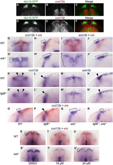
Fgf signaling regulates expression domains of chemokine pathway components. (A-F) The (A-C) cxcl12a and (D-F) cxcl12b genes are transcribed in regions adjacent to the dHb at 35 hpf, as shown by fluorescence in situ hybridization and immunolabeling for GFP in TgBAC(dbx1b:GFP) embryos. (G-H′) cxcl12b and (I-J′) ackr3b expression domains (brackets) are expanded in wls mutants. In fgf8a mutants, (K-L′) cxcl12b and (M-N′) ackr3b dorsal diencephalic expression domains (arrowheads) are reduced. Although the cxcl12b expression domain is reduced in (P) fgf8a mutants and expanded in (Q) wls mutants, (R) fgf8a;wls double mutants show a pattern more similar to (O) WT. Between 24 and 48 hpf, WT and wls mutant siblings were treated with either (S,S′) 0.3% DMSO, (T,T′) 18 µM SU5402+0.3% DMSO, or (U,U′) 24 µM SU5402+0.3% DMSO. Following treatment with 24 µM SU5402, cxcl12b expression in wls mutants is restored to the WT pattern (brackets). Scale bar: 30 µm. Refer to Table 1 for numerical values. Dorsal (A-G′,I,I′,K,K′,M,M′,S-U′) and lateral (H,H′,J,J′,L,L′,N,N′,O-R) views are shown. Scale bars: 50 µm. |
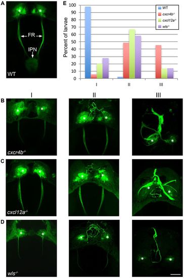
Chemokine signaling directs outgrowth of dHb axons. (A) In 5 dpf WT larvae, TgBAC(gng8:CAAX-GFP) labels neurons in the left and right dHb and their efferent axons in the FR, which terminate at the midbrain IPN. (B-D) Axonal morphology of 5 dpf (B) cxcr4b, (C) cxcl12a and (D) wls mutants falls into three general phenotypic classes: in Class I, the majority of axons fasciculate normally and project posteriorly; in Class II, some ectopic axons extend anterior to the dHb; and in Class III, the majority of axons grow anteriorly and fasciculate as a thick midline bundle. Asterisks indicate the dHb. Scale bar: 50 µm. (E) Percentage of WT siblings (blue, n=41) and cxcr4b (red, n=35), cxcl12a (green, n=30) and wls (purple, n=43) mutants with Class I, II or III axonal morphology. |

Chemokine signaling reporter labels newly born neurons and emerging axons. TgBAC(cxcr4b:cxcr4b-mKate2-IRES-GFP-CAAX) labeling at (A) 2 and (B) 3 dpf. GFP labels the membranes of dHb neurons and axons. The Cxcr4b chemokine receptor fusion protein (red) is found in neurons near the ventricular zone and on proximal (arrowheads) but not distal (arrows) regions of their axons. Scale bar: 50 µm. EXPRESSION / LABELING:
|
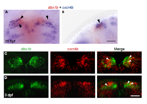
Partial overlap in cxcr4b and dbx1b expression. Visualization of dbx1b and cxcr4b transcripts by (A,B) colormetric in situ hybridization at 35 hpf or by (C,D) fluorescent in situ hybridization at 3 dpf. (A,C) Dorsal (B) lateral and (D) frontal views show partial co-localization of transcripts in a subset of dHb cells (arrowheads). Scale bar, 30 μm. EXPRESSION / LABELING:
|
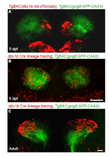
Medial dbx1b+ cells give rise to all dHb neurons. TgBAC(gng8:CAAX-GFP) labels the cell membranes of dHb neurons (green). (A) TgBAC(dbx1b:nls-dTomato) labels cells medial and ventral to the GFP labeled neurons. Dorsal view, 5 dpf. (B,C) Lineage tracing using Cre recombinase under control of the dbx1b promoter [TgBAC(dbx1b:Cre-mCherry)] and a floxed reporter line [Tg(β-actin:loxP-hmgb1-eCFP-loxP-H2B-mCherry)] labels neurons throughout the dHb with nuclear mCherry at (B) 5 dpf and (C) in adult brain sections. Scale bars are (A,B) 30 μm and (C) 50 μm. |
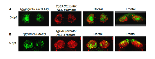
Persistence of fluorescent labeling in dHb neurons of transgenic larvae. In 5 dpf TgBAC(cxcr4b:nls-tdTomato) larvae, dTomato labeling persists in dHb neurons distinguished by (A) TgBAC(gng8:CAAX-GFP) or (B) Tg(HuC:H2B-GCaMP6s) labeling. Scale bar is 30 μm. |
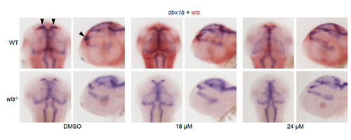
Inhibition of Fgf signaling reduces dbx1b and cxcl12b expression domains in a dose-dependent manner. Between 24 and 48 hpf, WT and wls mutant sibling embryos were treated with either 0.3% DMSO, 18 μM SU5402 + 0.3% DMSO, or 24 μM SU5402 + 0.3% DMSO. Diencephalic expression of dbx1b (arrowheads) is reduced in a dose-dependent manner in WT and wls mutants. |
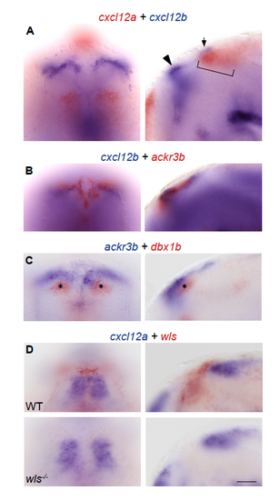
Diencephalic expression of genes in the Cxcr4-chemokine signaling pathway. (A) The cxcl12a gene is expressed ventral and caudal (bracket) to the developing habenulae. cxcl12b transcripts are found in high levels rostral (arrowhead) and lower levels caudal (arrow) to the dHb. (B) Transcripts for ackr3b are found anterior and medial to the rostral domain of cxcl12b expression and (C) the dbx1b+ progenitors (asterisks). (D) Expression of cxcl12a is not affected in wls mutants. Dorsal (left) and lateral (right) views. All embryos are 35 hpf. Scale bar is 50 μm. |