
|
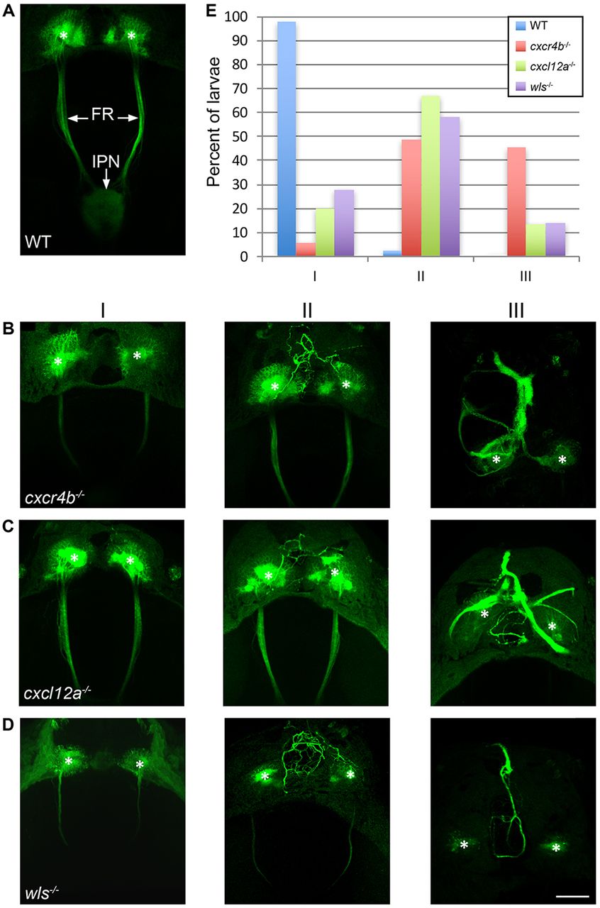
Fig. 7
Chemokine signaling directs outgrowth of dHb axons. (A) In 5 dpf WT larvae, TgBAC(gng8:CAAX-GFP) labels neurons in the left and right dHb and their efferent axons in the FR, which terminate at the midbrain IPN. (B-D) Axonal morphology of 5 dpf (B) cxcr4b, (C) cxcl12a and (D) wls mutants falls into three general phenotypic classes: in Class I, the majority of axons fasciculate normally and project posteriorly; in Class II, some ectopic axons extend anterior to the dHb; and in Class III, the majority of axons grow anteriorly and fasciculate as a thick midline bundle. Asterisks indicate the dHb. Scale bar: 50 µm. (E) Percentage of WT siblings (blue, n=41) and cxcr4b (red, n=35), cxcl12a (green, n=30) and wls (purple, n=43) mutants with Class I, II or III axonal morphology.