
- Title
-
Zebrafish Sox7 and Sox18 function together to control arterial-venous identity
- Authors
- Pendeville, H., Winandy, M., Manfroid, I., Nivelles, O., Motte, P., Pasque, V., Peers, B., Struman, I., Martial, J.A., and Voz, M.L.
- Source
- Full text @ Dev. Biol.

Spatio-temporal distribution of the zebrafish Sox7 and Sox18 transcripts from bud stage to 28 hpf. (A,F) Posterior views of bud stage embryos indicating that Sox7 and Sox18 are expressed in stripes corresponding to the LPM. (B,G) Expression of Sox7 and Sox18 in the LPM persists throughout somitogenesis. Staining occurs at the anterior and the posterior parts of 5 somite embryos. (C,H) At 18 somite stage, a signal corresponding to Sox7 and Sox18 is seen in the presumptive axial vessels. (D) At 24 hpf, Sox7 is mostly expressed in the DA, the head vessels, the ICM and in two stripes located in the hindbrain. Preferential expression of Sox7 in the dorsal aorta is confirmed in the close-up view of the midtrunk vasculature shown in panel E. (I) Sox18 transcripts can be detected in the head vasculature, the ICM, the dorsal aorta and the axial vein. (J) Close-up view of the trunk showing Sox18 expression in both axial vessels at 24 hpf. Lateral (C–E,H–J) or dorsal views (B,G). Embryos are shown with anterior to the left. The developmental stage is indicated in each panel. DA: dorsal aorta; AV: axial vein; NC: notochord; LPM: lateral plate mesoderm; ICM: intermediate cell mass. (K–M) Double fluorescent in situ for the vascular marker Flk1 and Sox7 on wild-type embryos at 18 somite stage. Confocal views of transverse vibratome sections show that Sox7+ positive cells totally overlap with Flk1+ cells. (N–P) Double staining of Scl and Sox7 show a population of Scl+-Sox7- cells located in the presumptive ICM (arrow in panel P). Panels M and P are merged views counterstained with Topro3 (blue). |
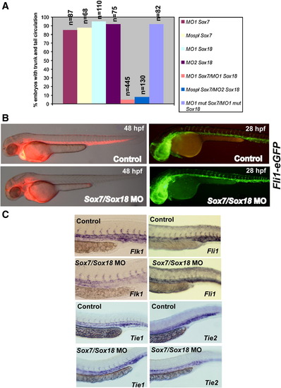
Knockdown of Sox7 together with Sox18 leads to a loss of circulation in the trunk and tail. (A) Targeting both Sox7 and Sox18 using different sets of morpholinos (4 ng of each) prevents trunk circulation in morphants. Sox7 or Sox18 knockdown alone shows no effect. (B) Left panel: Microangiography analyses to assess the functional integrity of the vasculature of 48 hpf control and Sox7/Sox18 morphants. The main axial vessels of double morphants embryos, injected with 4 ng of MO1 Sox18 and 4 ng of MO1 Sox7, show no uptake of the fluorescent dye whereas circulation in the head is not affected. Right panel: Normal Fli1 expression in Sox7/Sox18 knockdown embryos reveals no vascular apparent defect in the posterior vasculature at 28 hpf. (C) Lateral views of the trunk and tail, shown with anterior to the left, of embryos at 28 hpf. As evidenced by whole mount in situ with specific riboprobes, no significant difference in the vasculogenic expression of Fli1, Tie1, Tie2 is observed between control embryos and Sox7/Sox18 double morphants. There is a mild decrease in Flk1 expression at that particular stage but not at earlier time points (data not shown). |
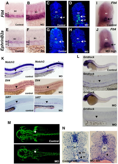
Sox7/Sox18 morphants display abnormal arterial–venous specification and arteriovenous shunts. Except in panels I,J, all embryos shown resulted of injection of regular doses of control or Sox7/Sox18 morpholinos (4 ng of each) and are fixed at 28 hpf. Pictures and sections are made at the trunk level. (A–D) In situ staining with the venous-specific marker Flt4 shows an ectopic expression in the dorsal aorta while the domain corresponding to the artery-specific marker EphrinB2a (panels E–H) is reduced. Panels C,D,G,H are confocal images of transverse sections of fluorescent embryos labeled by in situ. (I,J) Transverse sections of embryos injected with high doses of Sox7/Sox18 morpholinos (6 ng of each). As revealed by Flt4 expression, the vein is widely expanded, at the expense of the artery. (K) Expression of the artery markers Notch3, hRT and Dll4 is not affected in double morphants. (L) There is an abolition of Gridlock expression in the double morphants within the aorta while its expression in the brain was not affected. (M) White arrows mark the proximal aorta at the level where the paired lateral dorsal aortae normally fused into a single tube. 28 hpf Tg(fli1:EGFP)Y1 control fishes show well differentiated proximal aorta whereas morphants display strong dysmorphogenesis of the proximal aorta. (N) Hematoxylin–eosin stained sections reveal arteriovenous shunts in the trunk region in Sox7/Sox18 morphants. NC = notochord; DA = dorsal aorta; AV = axial vein. |

Sox7 and Sox18 are not involved in molecular pathways leading to the commitment of mesoderm to a blood fate. At early stages, formation of hematopoietic precursors appeared unaffected, as evident from normal Scl expression in the posterior LPM at 9 somite stage and from normal Gata1 and Scl expression at 18 somite stage in Sox7/Sox18 morphants. EXPRESSION / LABELING:
|

Influence of Shh, VEGF and Notch signaling on Sox7 and Sox18 expression. All embryos are shown at 24 hpf, and pictures were taken from lateral views at the trunk level. (A) Sox7 expression is lost in Smu mutant embryos and in embryos chemically impaired with cyclopamine whereas Sox18 expression is maintained. (B) Embryos treated with a VEGF receptor tyrosine kinase inhibitor show no expression of Sox7. Sox18 still appears expressed in these embryos. (C) Sox7 and Sox18 expression are maintained in Mindbomb mutants that are impaired in the Notch signalling. |
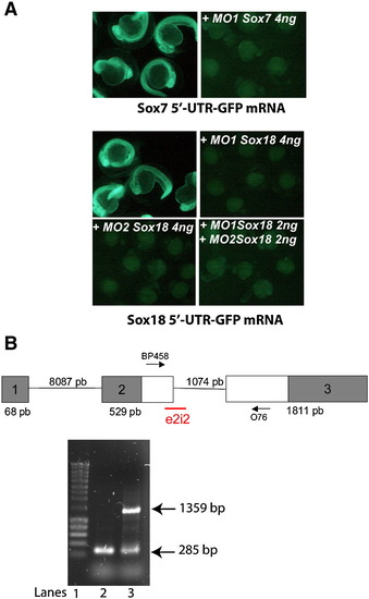
(A) Targeting efficiency of Sox 7 and Sox18 morpholinos. Messenger RNA synthesized from Sox7 and Sox18 5′UTR–GFP fusion constructs was injected in the embryos and re-injected afterwards with the respective morpholino. mRNA and morpholinos were not coinjected simultaneously, as in the test tube, the binding of the morpholino to its target is greatly favored compared to the in vivo situation in the embryo. If this binding is not totally and quickly reversible in the embryo, injecting together would lead to a reduction of the fluorescence which does not reflect the targeting efficiency of the morpholino in vivo. In all situation, GFP expression was totally abolished when MO1 Sox7, MO1 Sox18 or MO2 Sox18 were injected at a concentration of 4 ng/egg with 300 pg of the corresponding gene's 5′UTR–GFP expression construct. (B) Targeting efficiency of MOE2i2 Sox7 morpholino. Splice-site-targeted SOX7 morpholino alters splicing in zebrafish. Upper part: Genomic structure of zebrafish SOX7 gene. The ORF is indicated by white boxes. Splice site targeted by e2i2 is shown as well as the location of the primers used for the PCR. Lower part: Reverse transcriptase polymerase chain reaction (RT-PCR) analysis of Sox7 mRNA structure in control (lane 2) and e2i2 morphants (lane 3) using BP458 and O76. In the e2i2 morphants, we can observe a reduction of intensity of the band at 285 pb corresponding to the spliced transcript together with the apparition of a 1359 bp band corresponding to a non-spliced transcript containing the second intron. Lane 1 is the Smart ladder of Eurogentec (Belgium). |

Unillustrated author statements EXPRESSION / LABELING:
PHENOTYPE:
|
Reprinted from Developmental Biology, 317(2), Pendeville, H., Winandy, M., Manfroid, I., Nivelles, O., Motte, P., Pasque, V., Peers, B., Struman, I., Martial, J.A., and Voz, M.L., Zebrafish Sox7 and Sox18 function together to control arterial-venous identity, 405-416, Copyright (2008) with permission from Elsevier. Full text @ Dev. Biol.