
- Title
-
RUNX1-Evi-1 fusion gene inhibited differentiation and apoptosis in myelopoiesis: an in vivo study
- Authors
- Shen, L., Zhu, J., Chen, F., Lin, W., Cai, J., Zhong, J., Zhong, H.
- Source
- Full text @ BMC Cancer
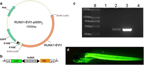
Generation of Tg(RE:HSE:EGFP) zebrafish line. (a) Schematic diagram of the structure of PSGH2/RUNX1-Evi-1 recombinant plasmid. A human-RUNX1-Evi-1 fragment was cloned into the EcoRI and EcoRV sites of the PSGH2 vector. (b) A schematic presentation of the eight multimerized heat shock element (HSE) promoter, which is flanked by two minimal promoters in opposed orientation (black arrowhead) to bidirectionally induce EGFP and RUNX1-Evi-1 expression. The vector is flanked by I-SceI meganuclease sites (arrows). pA, SV40 polyadenylation signal. (c) Transgenic verification by PCR: M: TAKARA DL2000 marker; lane 1 and 2: wild type and Tg(RE:HSE:EGFP) zebrafish larvae at 3 dpf, respectively; lane 3: PSGH2/RUNX1-Evi-1 plasmid; lane 4: double distilled water. (d) EGFP expression in Tg(RE:HSE:EGFP) zebrafish F2 generation at 3dpf (�4) EXPRESSION / LABELING:
|
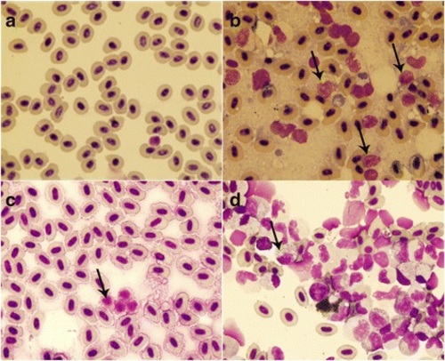
Cytological analysis of Tg(RE:HSE:EGFP) zebrafish. Cytology of hematopoietic cells from WT (a) and Tg(RE:HSE:EGFP) F2 generation (b) zebrafish at 60 dpf. The blood cells from WT fish were predominantly erythrocytes, and by contrast, erythrocytes were significantly inhibited in Tg(RE:HSE:EGFP) fish, enriched for abundant blast-like cells, which are larger than the erythrocytes and have high nuclear to cytoplasmic ratios, containing multiple large nucleoli (black arrow). These blasts were similar to that of human AML peripheral blood. The similar feature presented in single cell suspensions of kidneys from WT (c) and Tg F2 generation (d) PHENOTYPE:
|
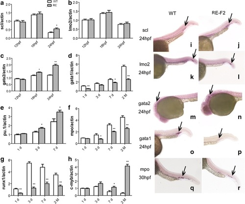
RUNX1-Evi-1 reprogrammed lineage-specific hematopoietic transcription factors. Scl (a), lmo2 (b) and gata2 (c) were detected by qRT-PCR in WT, and Tg(RE:HSE:EGFP) F2 generation embryos at 12 hpf, 18 hpf and 24 hpf. Gata1 (d), pu.1 (e), mpo (f), runx1(g), and c-myb (h) expressed in WT, Tg F2 zebrafish at 1 dpf, 3 dpf, 7 dpf and 60 dpf. Compared with WT, scl, gata2, pu.1, and c-myb were up-regulated, while gata1, mpo, and runx1 were down-regulated in Tg fish. In situ hybridization of scl (i-j), lmo2 (k-l), gata2 (m-n), and gata1 (o-p) at 24hpf and mpo (q-r) at 30hpf in WT and Tg F2 embryos demonstrated the same tendency. *P < 0.05; **P < 0.01 EXPRESSION / LABELING:
PHENOTYPE:
|

ZFIN is incorporating published figure images and captions as part of an ongoing project. Figures from some publications have not yet been curated, or are not available for display because of copyright restrictions. PHENOTYPE:
|
|
ZFIN is incorporating published figure images and captions as part of an ongoing project. Figures from some publications have not yet been curated, or are not available for display because of copyright restrictions. |
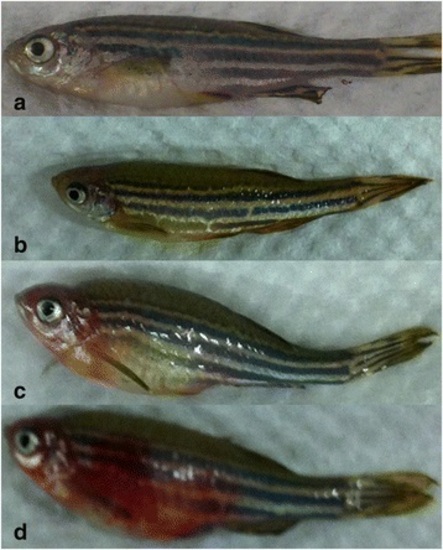
The appearance changes in Tg(RE:HSE:EGFP) adult zebrafish. (a) WT AB strain zebrafish; (b-d) various appearances with pathological changes presented in transgenic zebrafish, developmental delay (b), edema (c), and bleeding (d) PHENOTYPE:
|