
- Title
-
The transcription factor hairy/E(spl)-related 2 induces proliferation of neural progenitors and regulates neurogenesis and gliogenesis
- Authors
- Cheng, Y.C., Chiang, M.C., Shih, H.Y., Ma, T.L., Yeh, T.H., Huang, Y.C., Lin, C.Y., Lin, S.J.
- Source
- Full text @ Dev. Biol.

The expression of her2 in developing zebrafish embryos. (A) Schematic illustration of a gastrula stage embryo in lateral view. Prospective epidermis (epi) is green; prospective nervous system (neuro) is blue. an, animal pole; D, dorsal; V, ventral; ve, vegetal pole. (B-M) In situ hybridization analysis of the expression of her2 in the developing nervous system. (C-D) A coronal section of the her2-stained neural ectodermal region (as marked in B) with the double in situ hybridization for her2 (purple) and sox2 (red) and that shows overlapping expression. The expression of her2 was detected by NBT/BCIP and sox2 was detected by Fast Red. The stages of the embryos are shown in the bottom left corner of each panel. (B, E, G, I, K, and M) Dorsal views, with anterior to the left. (F, H, J, and L) Lateral view, anterior to the left. Arrows in E indicate her2 expressing cells in the proneuronal domains. The square bracket in J indicates the zona limitans intrathalamica (zli). Abbreviations: ce, cerebellum; dt, dorsal telencephalon; hb, hindbrain; mb, midbrain; mhb, midbrain-hindbrain boundary; psm, presomitic mesoderm; sc, spinal cord; tec, tectum; tel, telencephalon; th, thalamus; vt, ventral telencephalon; vpt, ventral-posterior telencephalon. EXPRESSION / LABELING:
|
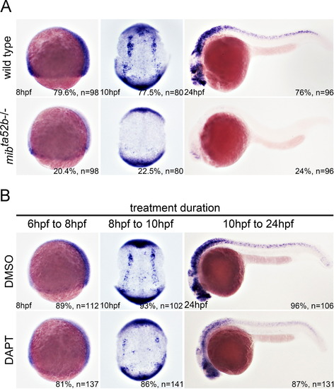
The expression of her2 is lost in Notch-deficient embryos. (A) The expression of her2 was analyzed in mibta52b homozygous mutants and wild-type siblings by in situ hybridization. The stages are shown in the bottom left corner. Left and right panels are lateral views, and the middle panels are dorsal views. The arrows indicate her2 expression in the neural progenitor cells. (B) DAPT treatment was performed at different time points, and the embryos were harvested at different stages as indicated, showing that DAPT treatment caused a downregulation of her2 expression. EXPRESSION / LABELING:
|

Knockdown of Her2 causes developmental abnormalities. (A) Embryos co-injected with 52her2-EGFP cRNA and control morpholino showed strong expression of EGFP (left panel). In contrast, the EGFP signal was abolished in embryos co-injected with 52her2-EGFP cRNA and her2 MO1 (right panel). (B) Representative images of morpholino-injected embryos. The asterisks denote edema in the fourth ventricle. Brain malformation is marked by arrows. These morphological phenotypes can be rescued by concomitant injection with 2 ng her2 cRNA. The bottom panels are the enlarged brain regions of the upper panels. Note that the structure of the midbrain-hindbrain boundary was unaffected by the her2 morpholino (arrowheads). |
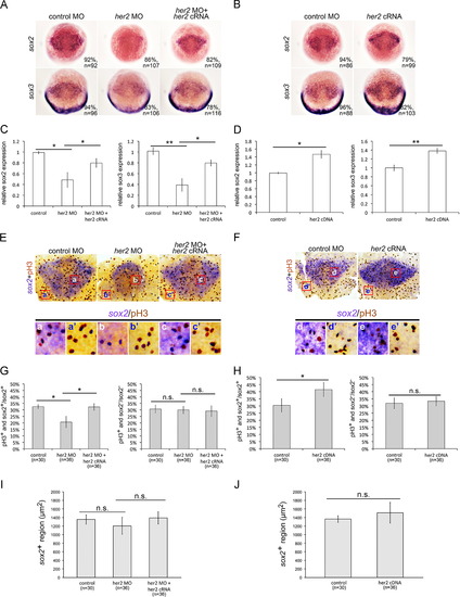
Her2 regulates neural progenitor proliferation. (A) The expression of neural progenitor markers sox2 and sox3 was downregulated in Her2-knockdown embryos and was rescued by co-injection with her2 cRNA. (B) her2 induced the expression of sox2 and sox3. (C and D) The results of in situ hybridization in A and B were quantitatively confirmed by qPCR analysis, respectively. (E, F) Proliferation of neural progenitors was detected by in situ hybridization using a sox2 riboprobe (purple) and counterstaining with phosphohistone H3 antibody (brown). The bottom panels are representative of the enlarged regions of the upper panels, as indicated. (G, H) The proportions of the phosphohistone H3- and sox2-positive cells among the total sox2-positive cells were quantified (left panels in G and H), showing that Her2 knockdown resulted in the decreased proliferation of sox2-positve neurons (left panel in G) and that her2 cRNA induced the proliferation of sox2-positive neurons (left panel in H). The right panels in G and H show the quantification of the proportions of phosphohistone H3-positive and sox2-negative cells in sox2-negative cells that were counted in the adjacent surface ectoderm, which showed no significant deviation in Her2-distorted embryos. (I, J) The sox2-positive region was quantified by ImageJ. All panels show embryos at 75% epiboly. pH3, phosphohistone H3; *, p<0.05; **, p<0.01; n.s., not significant. EXPRESSION / LABELING:
PHENOTYPE:
|

Knockdown of Her2 induces premature differentiation of neuronal precursors. (A) Injection of her2 morpholino downregulated the expression of neurog1, deltaA, ascl1a, and ascl1b and upregulated the expression of elavl3 at the bud stage. The phenotypes caused by morpholino injection could be rescued by concomitant injection with her2 cRNA. (B) qPCR analysis confirmed the results obtained by in situ hybridization. (C) Compared with the controls, injection of her2 cRNA did not cause a significant alteration in proneural markers (neurog1, deltaA, ascl1a, and ascl1b). (D) qPCR quantification of the results in C. **, p<0.01; n.s., not significant. |
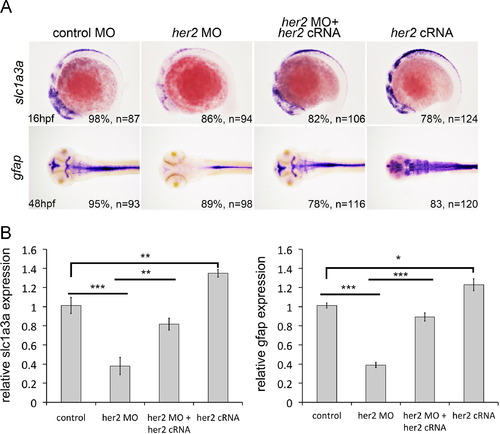
Her2 induces glial differentiation. (A) In situ hybridization showing the expression of slc1a3a and gfap was reduced in Her2-knockdown embryos, whereas injection of her2 cRNA upregulated their expression. The stages of the embryos are shown in the bottom-left corner. (B) qPCR analysis confirmed the results of in situ hybridization shown in A. *, p<0.05; **, p<0.01; ***, p<0.001. EXPRESSION / LABELING:
PHENOTYPE:
|
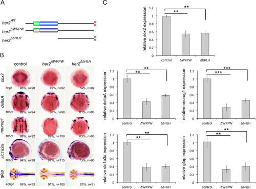
Resemblance between the phenotypes of her2ΔbHLH- or her2ΔWRPW-injected embryos and those of her2 morphants. (A) Schematic illustration of her2ΔbHLH and her2ΔWRPW deletion constructs. (B) In situ hybridization showing injection of either her2ΔbHLH or her2ΔWRPW was sufficient to downregulate markers for neural progenitors and neuronal and glial derivatives, as observed in Her2 morphants. The stages of the embryos are shown in the bottom- left corner. These results were confirmed by qPCR (C). *, p<0.05; **, p<0.01. EXPRESSION / LABELING:
|

Expression of her genes in proneuronal domains and interproneuronal domains. In situ hybridization that was performed on embryos at the bud stage shows that her2, her4, her12, and her15.1 are expressed in the proneuronal domains, whereas her3 is expressed in the interproneuronal domains. The interproneuronal domains are indicated by square brackets. |
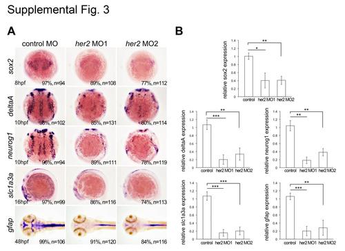
Embryos injected with MO1 or MO2 resulted in identical phenotypes. Injection of either MO1 or MO2 causes the same effects on markers for neural progenitors (sox2), neuronal precursors (deltaA and neurog1), glial precursors (slc1a3a), or radial glia (gfap), as shown by in situ hybridization (A) and quantified by qPCR (B). *, p < 0.05; **, p < 0.01. |
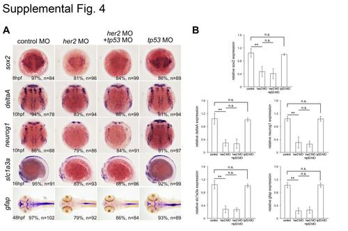
The phenotypes in Her2 morphants were not caused by nonspecific p53 activation. Embryos co-injected with her2 MO and tp53 MO were compared with Her2 morphants and did not cause any detectable deviation from all markers tested, and injection of tp53 MO alone did not show any significant alterations compared to the controls, as shown by in situ hybridization (A) and quantified by qPCR (B).*, p < 0.05; **, p < 0.01; n. s., not significant. |

Her2 does not regulate neural patterning or neural versus epidermal fate determination. (A) Schematic illustration of a gastrula stage embryo in lateral view. Prospective tp63-positive epidermis is green; prospective otx2-positive and zic2b-positive neural ectoderm is magenta and blue, respectively. an, animal pole; D, dorsal; V, ventral; ve, vegetal pole. (B) The expression of otx2 and zic2b were downregulated by her2 MO and upregulated by her2 cRNA injections, as shown by in situ hybridization. (C) qPCR quantification for (B). (D) Quantification of the size of the otx2 or zic2b expression domain, showing no significant alterations from her2 cRNA or her2 MO injections. Alterations in Her2 expression had no significant effects on the expression of tp63. Region of interest was quantified by ImageJ *, p<0.05; **, p<0.01; n.s., not significant. |
Reprinted from Developmental Biology, 397(1), Cheng, Y.C., Chiang, M.C., Shih, H.Y., Ma, T.L., Yeh, T.H., Huang, Y.C., Lin, C.Y., Lin, S.J., The transcription factor hairy/E(spl)-related 2 induces proliferation of neural progenitors and regulates neurogenesis and gliogenesis, 116-28, Copyright (2015) with permission from Elsevier. Full text @ Dev. Biol.