FIGURE SUMMARY
Title

Disruption of sirtuin 7 in zebrafish facilitates hypoxia tolerance

Authors
Liao, Q., Zhu, C., Sun, X., Wang, Z., Chen, X., Deng, H., Tang, J., Jia, S., Liu, W., Xiao, W., Liu, X.
Source
Full text @ J. Biol. Chem.

Zebrafish sirt7 is suppressed by hypoxia.A, quantitative real-time PCR analysis (qPCR) of sirt7, as well as well-defined hypoxia-responsive genes, including cited2, pai1, phd3, vegfaa, and epoa, in zebrafish larvae (3 dpf) under normoxia (Nor) and hypoxia (Hyp). B, qPCR analysis of sirt7, as well as well-defined hypoxia-responsive genes, including phd3, cited2, ldha, and vegfaa, in ZFL cells under normoxia (Nor) and hypoxia (Hyp). C, qPCR analysis of sirt7 in ZFL cells transfected with empty vector control (Flag empty) or Flag-hif-1αa, or Flag-hif-1αb, or Flag-hif-2αa, or Flag-hif-2αb under normoxia. D, qPCR analysis of phd3 in ZFL cells transfected with empty vector control (Flag empty) or Flag-hif-1αa, or Flag-hif-1αb, or Flag-hif-2αa, or Flag-hif-2αb under normoxia. E, qPCR analysis of cited2 in ZFL cells transfected with empty vector control (Flag empty) or Flag-hif-1αa, or Flag-hif-1αb, or Flag-hif-2αa, or Flag-hif-2αb under normoxia. F, qPCR analysis of ldha in ZFL cells transfected with empty vector control (Flag empty) or Flag-hif-1αa, or Flag-hif-1αb, or Flag-hif-2αa, or Flag-hif-2αb under normoxia. G, qPCR analysis of vegfaa in ZFL cells transfected with empty vector control (Flag empty) or Flag-hif-1αa, or Flag-hif-1αb, or Flag-hif-2αa, or Flag-hif-2αb under normoxia. p Values were calculated by unpaired Student's t test (A and B) or two-way ANOVA analysis (CG); ns, not significant, ∗p < 0.05, ∗∗p < 0.01, ∗∗∗p < 0.001 and ∗∗∗∗p < 0.0001; data based on one representative experiment performed in three biological replicates from at least three independent experiments (mean ± SD).

EXPRESSION / LABELING:
Genes:
Fish:
Condition:
Anatomical Term:
Stage: Protruding-mouth
PHENOTYPE:
Fish:
Condition:
Observed In:
Stage: Protruding-mouth

sirt7-null zebrafish larvae (sirt7ihblqs4/ihblqs4) are more tolerant to hypoxia.A, Western blot analysis of sirt7 expression in sirt7-null mutant (sirt7ihblqs4/ihblqs4) and the wildtype sibling (sirt7+/+) (3 dpf). B, quantitative real-time PCR (qPCR) analysis of phd3 mRNA in sirt7-null mutant (sirt7ihblqs4/ihblqs4) and the wildtype sibling (sirt7+/+) (3 dpf) under normoxia (Nor) and hypoxia (Hyp). C, qPCR analysis of pai1 mRNA in sirt7-null mutant (sirt7ihblqs4/ihblqs4) and the wildtype sibling (sirt7+/+) (3 dpf) under normoxia (Nor) and hypoxia (Hyp). D, qPCR analysis of cited2 mRNA in sirt7-null mutant (sirt7ihblqs4/ihblqs4) and the wildtype sibling (sirt7+/+) (3 dpf) under normoxia (Nor) and hypoxia (Hyp). E, qPCR analysis of vegfaa mRNA in sirt7-null mutant (sirt7ihblqs4/ihblqs4) and the wildtype sibling (sirt7+/+) (3 dpf) under normoxia (Nor) and hypoxia (Hyp). F, qPCR analysis of epoa mRNA in sirt7-null mutant (sirt7ihblqs4/ihblqs4) and the wildtype sibling (sirt7+/+) (3 dpf) under normoxia (Nor) and hypoxia (Hyp). G, o-dianisidine staining of erythrocytes in sirt7-null mutant (sirt7ihblqs4/ihblqs4) and the wildtype sibling (sirt7+/+) (6 dpf) under normoxia (Nor) and hypoxia (Hyp). H, quantitation of erythrocytes in (G) as determined by normalizing the intensities of o-dianisidine-stained cells. I, photographs of sirt7-null mutant (sirt7ihblqs4/ihblqs4) and the wildtype sibling (sirt7+/+) (3 dpf) under normoxia (Nor) and hypoxia (Hyp). J, survival curve of sirt7-null mutant (sirt7ihblqs4/ihblqs4) and the wildtype sibling (sirt7+/+) (3 dpf) under normoxia (Nor) and hypoxia (Hyp). p Values were calculated by two-way ANOVA analysis (B, C, D, E, F, and H) or log-rank test (J); ∗∗p < 0.01, ∗∗∗p < 0.001, and ∗∗∗∗p < 0.0001; data based on one representative experiment performed in three biological replicates from at least three independent experiments (mean ± SD).

sirt7-null zebrafish larvae (sirt7ihblqs7/ihblqs7) are more tolerant to hypoxia.A, Western blot analysis of sirt7 expression in sirt7-null mutant (sirt7ihblqs7/ihblqs7) and the wildtype sibling (sirt7+/+) (3 dpf). B, quantitative real-time PCR (qPCR) analysis of phd3 mRNA in sirt7-null mutant (sirt7ihblqs7/ihblqs7) and the wildtype sibling (sirt7+/+) (3 dpf) under normoxia (Nor) and hypoxia (Hyp). C, qPCR analysis of pai1 mRNA in sirt7-null mutant (sirt7ihblqs7/ihblqs7) and the wildtype sibling (sirt7+/+) (3 dpf) under normoxia (Nor) and hypoxia (Hyp). D, qPCR analysis of cited2 mRNA in sirt7-null mutant (sirt7ihblqs7/ihblqs7) and the wildtype sibling (sirt7+/+) (3 dpf) under normoxia (Nor) and hypoxia (Hyp). E, qPCR analysis of vegfaa mRNA in sirt7-null mutant (sirt7ihblqs7/ihblqs7) and the wildtype sibling (sirt7+/+) (3 dpf) under normoxia (Nor) and hypoxia (Hyp). F, qPCR analysis of epoa mRNA in sirt7-null mutant (sirt7ihblqs7/ihblqs7) and the wildtype sibling (sirt7+/+) (3 dpf) under normoxia (Nor) and hypoxia (Hyp). G, o-dianisidine staining of erythrocytes in sirt7-null mutant (sirt7ihblqs7/ihblqs7) and the wildtype sibling (sirt7+/+) (6 dpf) under normoxia (Nor) and hypoxia (Hyp). H, quantitation of erythrocytes in (G) as determined by normalizing the intensities of o-dianisidine-stained cells. I, photographs of sirt7-null mutant (sirt7ihblqs7/ihblqs7) and the wildtype sibling (sirt7+/+) (3 dpf) under normoxia (Nor) and hypoxia (Hyp). J, survival curve of sirt7-null mutant (sirt7ihblqs7/ihblqs7) and the wildtype sibling (sirt7+/+) (3 dpf) under normoxia (Nor) and hypoxia (Hyp). p Values were calculated by two-way ANOVA analysis (B, C, D, E, F, and H) or log-rank test (J); ∗∗p < 0.01, ∗∗∗p < 0.001, and ∗∗∗∗p < 0.0001; data based on one representative experiment performed in three biological replicates from at least three independent experiments (mean ± SD).

Zebrafish sirt7 suppresses hypoxia-responsive gene expression.A, Western blot analysis of indicated protein levels in EPC cells transfected empty vector control (Myc empty) or Myc-tagged zebrafish sirt7 (Myc-sirt7). B, luciferase activity of hypoxia-responsive element (HRE)-luciferase reporter in EPC cells transfected with empty vector control (Myc empty) or Myc-tagged zebrafish sirt7 (Myc-sirt7) under normoxia (Nor) or hypoxia (Hyp). C, Western blot analysis of indicated protein levels in ZFL cells transfected empty vector control (Myc empty) or Myc-tagged zebrafish sirt7 (Myc-sirt7). D, quantitative real-time PCR (qPCR) analysis of phd3 in ZFL cells transfected with empty vector control (Myc empty) or Myc-tagged zebrafish sirt7 (Myc-sirt7) under normoxia (Nor) or hypoxia (Hyp). E, qPCR analysis of vegfaa in ZFL cells transfected with empty vector control (Myc empty) or Myc-tagged zebrafish sirt7 (Myc-sirt7) under normoxia (Nor) or hypoxia (Hyp). F, qPCR analysis of ldha in ZFL cells transfected with empty vector control (Myc empty) or Myc-tagged zebrafish sirt7 (Myc-sirt7) under normoxia (Nor) or hypoxia (Hyp). G, qPCR analysis of cited2 in ZFL cells transfected with empty vector control (Myc empty) or Myc-tagged zebrafish sirt7 under normoxia (Nor) or hypoxia (Hyp). p Values were calculated by two-way ANOVA analysis (B, D, E, F, and G); ∗∗∗∗p < 0.0001; data based on one representative experiment performed in three biological replicates from at least three independent experiments (mean ± SD).

Zebrafish sirt7 binds to hif-1α and hif-2α.A, coimmunoprecipitation analysis of Myc-sirt7 with Flag-hif-1αa, or Flag-hif-1αb, or Flag-hif-2αa, or Flag-hif-2αb. HEK293T cells were transfected with Myc-sirt7, together with Flag empty vector, or Flag-hif-1αa, or Flag-hif-1αb, or Flag-hif-2αa, or Flag-hif-2αb. Anti-Flag-conjugated agarose beads were used for coimmunoprecipitation, and the indicated antibodies were used for detection. B, coimmunoprecipitation analysis of Myc-sirt7 with Flag-hif-1αa under normoxia (Nor) or hypoxia (Hyp). HEK293T cells were transfected with Myc-sirt7, together with Flag empty vector or Flag-hif-1αa, and cultured under normoxia (Nor) or hypoxia (Hyp) for 4 h. Anti-Flag-conjugated agarose beads were used for coimmunoprecipitation, and the indicated antibodies were used for detection. The Flag-hif-1αa protein under hypoxia was adjusted to be similar to that under normoxia. The immunoprecipitated Myc-sirt7 protein by Flag-hif-1αa in IP (∗) over the Myc-sirt7 protein in TCL (#) was determined (∗/#). C, coimmunoprecipitation analysis of Myc-sirt7 with Flag-hif-1αb under normoxia (Nor) or hypoxia (Hyp). HEK293T cells were transfected with Myc-sirt7, together with Flag empty vector or Flag-hif-1αb, and cultured under normoxia (Nor) or hypoxia (Hyp) for 4 h. Anti-Flag-conjugated agarose beads were used for coimmunoprecipitation, and the indicated antibodies were used for detection. The Flag-hif-1αb protein under hypoxia was adjusted to be similar to that under normoxia. The immunoprecipitated Myc-sirt7 protein by Flag-hif-1αb in IP (∗) over the Myc-sirt7 protein in TCL (#) was determined (∗/#). D, coimmunoprecipitation analysis of Myc-sirt7 with Flag-hif-2αa under normoxia (Nor) or hypoxia (Hyp). HEK293T cells were transfected with Myc-sirt7, together with Flag empty vector or Flag-hif-2αa, and cultured under normoxia (Nor) or hypoxia (Hyp) for 4 h. Anti-Flag-conjugated agarose beads were used for coimmunoprecipitation, and the indicated antibodies were used for detection. The Flag-hif-2αa protein under hypoxia was adjusted to be similar to that under normoxia. The immunoprecipitated Myc-sirt7 protein by Flag-hif-2αa in IP (∗) over the Myc-sirt7 protein in TCL (#) was determined (∗/#). E, coimmunoprecipitation analysis of Myc-sirt7 with Flag-hif-2αb under normoxia (Nor) or hypoxia (Hyp). HEK293T cells were transfected with Myc-sirt7, together with Flag empty vector or Flag-hif-2αb, and cultured under normoxia (Nor) or hypoxia (Hyp) for 4 h. Anti-Flag-conjugated agarose beads were used for coimmunoprecipitation, and the indicated antibodies were used for detection. The Flag-hif-2αb protein under hypoxia was adjusted to be similar to that under normoxia. The immunoprecipitated Myc-sirt7 protein by Flag-hif-2αb in IP (∗) over the Myc-sirt7 protein in TCL (#) was determined (∗/#). IP, immunoprecipitation; TCL, total cell lysates.

Zebrafish sirt7 promotes degradation of hif-1αa, hif-1αb, hif-2αa, and hif-2αb.A, Western blot analysis of hif-1αa expression in HEK293T cells transfected with Flag-hif-1αa together with an increased amount of Myc-tagged sirt7. B, Western blot analysis of hif-1αb expression in HEK293T cells transfected with Flag-hif-1αb together with an increased amount of Myc-tagged sirt7. C, Western blot analysis of hif-2αa expression in HEK293T cells transfected with Flag-hif-2αa together with an increased amount of Myc-tagged sirt7. D, Western blot analysis of hif-2αb expression in HEK293T cells transfected with Flag-hif-2αb together with an increased amount of Myc-tagged sirt7. E, Western blot analysis of indicated protein levels in sirt7-null mutant (sirt7ihblqs4/ihblqs4) and the wildtype sibling (sirt7+/+) (3 dpf). F, Western blot analysis of indicated protein levels in sirt7-null mutant (sirt7ihblqs7/ihblqs7) and the wildtype sibling (sirt7+/+) (3 dpf).

Zebrafish sirt7 facilitates ubiquitination of hif-1αa, hif-1αb, hif-2αa, and hif-2αb.AD, quantitative real-time PCR (qPCR) analysis of the mRNA levels of hif-1αa (A), hif-1αb (B), hif-2αa (C), or hif-2αb (D) in sirt7-null mutant (sirt7ihblqs4/ihblqs4) and the wildtype sibling (sirt7+/+) (3 dpf) under normoxia (Nor) and hypoxia (Hyp). EH, qPCR analysis of the mRNA levels of hif-1αa (E), hif-1αb (F), hif-2αa (G), or hif-2αb (H) in sirt7-null mutant (sirt7ihblqs7/ihblqs7) and the wildtype sibling (sirt7+/+) (3 dpf) under normoxia (Nor) and hypoxia (Hyp). IL, ubiquitination analysis of hif-1αa (I), hif-1αb (J), hif-2αa (K), or hif-2αb (L) in HEK293T cells transfected with indicated plasmids for 24 h and then treated with MG132 (20 μM) for 8 h. p Values were calculated by two-way ANOVA analysis (AH); ns, not significant; data based on one representative experiment performed in three biological replicates from at least three independent experiments (mean ± SD).

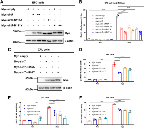
Zebrafish sirt7 attenuates hypoxia signaling independent of its deacetylase activity.A, Western blot analysis of indicated protein levels in EPC cells transfected with empty vector, wildtype sirt7, or the enzyme-deficient mutants sirt7-S115A and sirt7-H191Y. B, luciferase activity of HRE-luciferase reporter in EPC cells transfected with empty vector, wildtype sirt7, or the enzyme-deficient mutants sirt7-S115A and sirt7-H191Y under normoxia (Nor) or hypoxia (Hyp). C, Western blot analysis of indicated protein levels in ZFL cells transfected with empty vector, wildtype sirt7, or the enzyme-deficient mutants sirt7-S115A and sirt7-H191Y. D, quantitative real-time PCR (qPCR) analysis of phd3 in ZFL cells transfected with empty vector, wildtype sirt7, or the enzyme-deficient mutants sirt7-S115A and sirt7-H191Y under normoxia (Nor) or hypoxia (Hyp). E, qPCR analysis of cited2 in ZFL cells transfected with empty vector, wildtype sirt7, or the enzyme-deficient mutants sirt7-S115A and sirt7-H191Y under normoxia (Nor) or hypoxia (Hyp). F, qPCR analysis of ldha in ZFL cells transfected with empty vector, wildtype sirt7, or the enzyme-deficient mutants sirt7-S115A and sirt7-H191Y under normoxia (Nor) or hypoxia (Hyp). p Values were calculated by two-way ANOVA analysis (B, D, E, and F); ∗∗p < 0.01, ∗∗∗p < 0.001 and ∗∗∗∗p < 0.0001; data based on one representative experiment performed in three biological replicates from at least three independent experiments (mean ± SD).

Acknowledgments
This image is the copyrighted work of the attributed author or publisher, and ZFIN has permission only to display this image to its users. Additional permissions should be obtained from the applicable author or publisher of the image. Full text @ J. Biol. Chem.