
Fig. 6
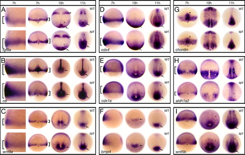
Components of the Fgf and Wnt pathway are more highly expressed in spt mutants. Expression of (A) fgf8a; (B) ntl; (C) wnt8a; (D) cdx4; (E) cdx1a; (F) bmp4; (G) chordin; (H) aldh1a2; and (I) wnt5b at: 7 h, 10 h and 11 h. Embryos at 7 h are shown in (A–C) a high magnification left side view and a low magnification dorsal view, (D–F) a left side view, and (G–I) a dorsal view. Older embryos are all shown in a dorsal posterior view. Designations (7 h): brackets, more or less expression; open circles, dorsal side; arrowhead, prechordal plate; (later stages): arrow, more expression in the posterior region and tailbud; asterisk, less expression in the paraxial mesoderm; arrowhead, more expression in the future midbrain-hindbrain boundary; open arrowhead, more expression in the intermediate mesoderm. |
| Genes: | |
|---|---|
| Fish: | |
| Anatomical Terms: | |
| Stage Range: | Shield to 1-4 somites |
Reprinted from Developmental Biology, 383(1), Warga, R.M., Mueller, R.L., Ho, R.K., and Kane, D.A., Zebrafish Tbx16 regulates intermediate mesoderm cell fate by attenuating Fgf activity, 75-89, Copyright (2013) with permission from Elsevier. Full text @ Dev. Biol.