FIGURE

Fig. 2

ID
ZDB-FIG-131213-49
Publication
Warga et al., 2013 - Zebrafish Tbx16 regulates intermediate mesoderm cell fate by attenuating Fgf activity
Other Figures
All Figure Page
Back to All Figure Page
Fig. 2

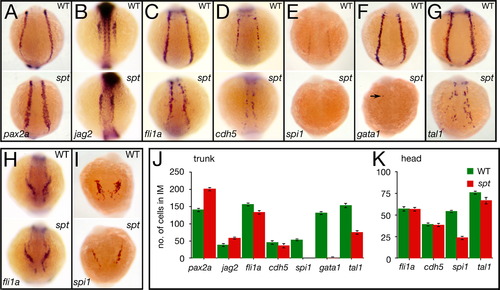
spt mutants possess more nephric precursors, but have fewer endothelial and hematopoietic precursors. (A–G) Expression in the trunk at 14.5 h of: (A) pax2a and (B) jag2, in nephric cells; (C) fli1a and (D) cdh5, in endothelial cells; and (E) spi1, in white, (F) gata1, in red, and (G) tal1, in all blood cells (arrow designates a single cell). (H, I) Expression in the head at 14.5 h of: (H) fli1a, in endothelial cells; and (I) spi1, in macrophages. (J, K) Quantification of intermediate mesoderm precursors for: (J) trunk, and (K) head. Graphs show the average number of cells per marker including standard error; 9 embryos for each category were counted. Embryos are shown from a (A–G) posterior view, or (H, I) anterior view with dorsal to the top.

Expression Data
Genes:
Fish:
Anatomical Terms:
Stage: 10-13 somites

Expression Detail
Antibody Labeling
Phenotype Data
Fish:
Observed In:
Stage: 10-13 somites

Phenotype Detail
Acknowledgments
This image is the copyrighted work of the attributed author or publisher, and ZFIN has permission only to display this image to its users. Additional permissions should be obtained from the applicable author or publisher of the image.

Reprinted from Developmental Biology, 383(1), Warga, R.M., Mueller, R.L., Ho, R.K., and Kane, D.A., Zebrafish Tbx16 regulates intermediate mesoderm cell fate by attenuating Fgf activity, 75-89, Copyright (2013) with permission from Elsevier. Full text @ Dev. Biol.