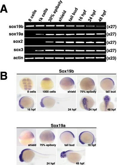
Expression of the SoxB1 family of genes during zebrafish embryonic development. a RT-PCR was used to analyse the expression level of the SoxB1 family of genes during zebrafish developmental stages, ranging from 8 cells to 48 hpf. b Expression analysis of Sox19b and Sox19a in zebrafish was determined by whole-mount in situ hybridization at the indicated stages (from 8 cells to 48 hpf)

Zebrafish embryos displayed NTDs after knockdown of Sox19b.a Western blot confirmed that Sox19b MO could interfere Sox19b mRNA translation. b The neural tube development of zebrafish embryos was abnormal after injection with a Sox19b MO at 48 hpf. There were two main results. The mild phenotype consisted of a smaller head and shorter body axis. The severe phenotype had a partial head area deletion, microphthalmos, shorter body axis, and tail turned up. Bar graphs show the statistical data for the embryo numbers. Data represent the mean of at least three independent experiments ± SD. **P < 0.01 versus control. c The zebrafish embryos developed to 24 hpf and 30 hpf, and the embryos injected with Sox19b MO showed that the area of the telencephalon and diencephalon decreased significantly compared with controls. The bar graphs show the percentage of normal embryos in each group. Data represent the mean of at least three independent experiments ± SD. **P < 0.01 versus control. d Whole-mount in situ hybridization analysis of neuroectoderm marker expression at 24 hpf. The expression of otx2 and pax2a were decreased in the Sox19b MO group compared with what was observed in the controls. Bar graphs show the percentage of normal embryos in each group. Data represent the mean of at least three independent experiments ± SD. *P < 0.05 versus control

Effect of Sox19b on NSCs in neural tubes. a H&E histological sections of the brain and spinal cord of zebrafish embryos at 24 hpf showed that the number of cells and cell layers in the neural tube decreased, and the ventricle expanded in the Sox19b MO-injected embryos compared with controls. b BrdU labelling of control MO- and Sox19b MO-injected embryos at 24 hpf. Data represent the mean of at least three independent experiments ± SD. **P < 0.01 versus control. c The expression of PCNA was detected by whole-mount in situ hybridization, and the expression was used to detect proliferating cells in zebrafish embryos of control MO MO- and Sox19b MO-injected embryos. d Immunolabelling of HuC (early neuronal marker) at 24 hpf showed that the expression of HuC was ectopic in Sox19b morphants, and the expression of HuC appeared in some regions that were enriched with NSCs. Data represent the mean of at least three independent experiments ± SD. *P < 0.05 versus control

NSCs in embryos injected with sox19b MO were prematurely differentiated. a Quantitative RT-PCR analysis for Ngn1, ascl1a, HuC, lslet1, and her3. Compared with the control group, the mRNA levels of Ngn1, ascl1a, HuC, and lslet1 in Sox19b MO-injection embryos increased significantly, but the mRNA level of her3 decreased significantly. b The expression levels of Ngn1, HuC, and lslet1 were detected by whole-mount in situ hybridization. Bar graphs show the percentage of normal embryos in each group. Data represent the mean of at least three independent experiments ± SD. *P < 0.05, **P < 0.01 versus control. c Quantitative RT-PCR analysis is shown for p21, p27, and p57. Knockdown of Sox19b caused a significant increase in the mRNA levels of cyclin-dependent kinase inhibitor. Data represent the mean of at least three independent experiments ± SD. *P < 0.05 versus control. d Control and Sox19b MO-injected embryos were analysed by immunofluorescence staining of PHH3 (a marker of mitosis, red). Data represent the mean of at least three independent experiments ± SD. *P < 0.05 versus control

The FGF pathway did not play the most important role in the Sox19b regulation of NSCs. a Quantitative RT-PCR analysis is shown for fgf3, fgf8, and er81. Compared with the controls, the mRNA levels of fgf3 and er81 in Sox19b MO-injected embryos decreased significantly. Data represent the mean of at least three independent experiments ± SD. *P < 0.05 versus control. b Compared with embryos injected with Sox19b MO, the NTD appearance in zebrafish embryos was not rescued by the addition of fgf3 mRNA. c Quantitative RT-PCR analysis for Ngn1, Huc, and Islet1. Fgf3 mRNA could not change the increase in Ngn1, Huc, or lslet1 induced by Sox19b MO treatment. Data represent the mean of at least three independent experiments ± SD. *P < 0.05, **P < 0.01 versus control; ns, no significant difference

EXPRESSION / LABELING:
Genes:
Fish:
Knockdown Reagents:
Anatomical Term:
Stage Range: Bud to 1-4 somites
PHENOTYPE:
Fish:
Knockdown Reagents:
Observed In:
Stage Range: Bud to Long-pec

Sox19b regulated NSCs through an epigenetic mechanism. a Western blot analysis of acetyl-H3 protein levels in control and Sox19b MO-injected embryos. Sox19b had no effect on the acetyl-H3 levels. b Western blot analysis of H3K27me3 and H3K9me3 levels in control and Sox19b MO-injected embryos. The H3K27me3 level decreased after knocking down sox19b, but the level of H3K9me3 remained unchanged. Data represent the mean of at least three independent experiments ± SD. *P < 0.05 versus control. c, d Control and Sox19b MO-injected embryos were immunoprecipitated with anti-H3K27me3 antibody, and the isolated DNA was analysed by using gene-specific ChIP primers. Rabbit IgG was used as a negative control. DNA from each ChIP sample was normalized by the corresponding input sample. Compared with the controls, the level of H3K27me3 in the Ngn1 and ascl1a promoter regions decreased significantly in Sox19b MO-injected embryos. Data represent the mean of at least three independent experiments ± SD. *P < 0.05 versus control

Sox19b regulated NSCs through EZH2. a Quantitative RT-PCR analysis is shown for EZH2. Compared with the controls, the mRNA level of EZH2 in Sox19b MO-injected embryos decreased significantly. b Western blot analysis of EZH2 protein levels in control and Sox19b MO-injected embryos. The protein level of EZH2 decreased after knocking down of Sox19b. Data represent the mean of at least three independent experiments ± SD. **P < 0.01 versus control

EXPRESSION / LABELING:
Gene:
Fish:
Knockdown Reagents:
Anatomical Term:
Stage Range: Bud to 1-4 somites
PHENOTYPE:
Fish:
Knockdown Reagents:
Observed In:
Stage Range: Bud to 1-4 somites

Schematic representation of Sox19b-regulated neural differentiation of NSCs in zebrafish neural tube development

Acknowledgments
This image is the copyrighted work of the attributed author or publisher, and ZFIN has permission only to display this image to its users. Additional permissions should be obtained from the applicable author or publisher of the image. Full text @ Stem Cell Res. Ther.