
- Title
-
Twisted gastrulation promotes BMP signaling in zebrafish dorsal-ventral axial patterning
- Authors
- Little, S.C., and Mullins, M.C.
- Source
- Full text @ Development
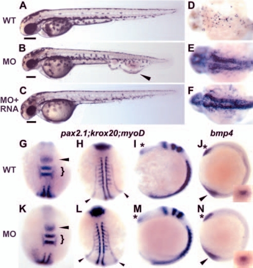
Low-level tsg1 knockdown causes a ventral tail vein edema and cell death in anterior structures, but does not alter patterning during early somitogenesis. Uninjected embryo (A); a wild-type embryo injected with 8 ng MO1 (B) displays a slightly reduced head and a large edema in, or dilation of, the ventral tail vein (arrowhead), in which the blood accumulates and circulation slows. (C) The vein edema can be rescued by injection of tsg1 mRNA (10% with edema phenotype, n=133, not shown) or tsg2 mRNA (8% with edema phenotype, n=106), whereas the reduction in head size is unaltered. Scale bars in A-C show the diameter of the eye as a measure of the extent of anterior tissues. TUNEL analysis as a measure of cell death in 24 hours post fertilization (hpf) uninjected wild-type (D) and 8 ng MO1-injected sibling (E) embryos. Cell death is not rescued by co-injection of tsg mRNA (F). (D-F) Anterior towards the left, dorsal view posterior to the MHB boundary. (G-N) Whole-mount in situ hybridization of uninjected embryos (G-J) or embryos injected with 8 ng MO1 (K-N). (G,K) Expression of pax2.1 (arrowhead) and krox20 (bracket) in the mid/hindbrain (MHB) boundary and rhombomeres 3 and 5, respectively. (H,L) More posterior views of the embryos in G and K. pax2.1 expression in the pronephric system (arrowheads) and myoD in anterior somitic mesoderm is unchanged. (I,M) Lateral views of untreated or injected embryos showing no alterations in anterior expression of pax2.1 in the eye field (asterisk). (J,N) Expression of bmp4 anteriorly (asterisk) and in the tail bud (arrowhead) at the three-somite stage is normal in injected embryos. Insets show tail bud views. (G-I,K-M) Eight somites. (J,N) Three somites. (G,H,K,L) Dorsal views with anterior upwards. (I,J,M,N) Lateral views, anterior is upwards. EXPRESSION / LABELING:
|

High level tsg1 knockdown dorsalizes the embryo. Uninjected embryos (A-E,K-M) compared with siblings injected with 25 ng MO5 (F-J,N,P,Q) or 32 ng MO1 (O). (A-J) Whole-mount in situ hybridization at 80% epiboly (mid-gastrulation); animal pole views, dorsal towards the right. Reduced ventral domain of eve1 (n=32/54), dlx3 (n=25/41) and gata2 (n=28/50) expression (A-C,F-H) and expanded foxb1.2 (n=34/46) and chordin (chd) (n=16/30) expression in dorsal regions (D,E,I,J) in injected compared with uninjected embryos (delineated by arrowheads) was observed. gsc expression in the dorsal midline prechordal plate mesoderm is not affected (A,F), similar to results in other dorsalized BMP signaling pathway mutants in zebrafish (Mullins et al., 1996; Nguyen et al., 1998). (K-Q) During somitogenesis, injected embryos display dorsalized characteristics similar to class 3 (N) and class 4 (O) dorsalized mutants, as assessed by the lateral extent of somites in live embryos (arrowheads in K,N,O), by whole-mount in situ hybridization with myod in anterior somitic mesoderm (M,P,Q), and by expansion of pax2.1 (arrowhead) and krox20 (bracket) expression in the MHB and rhombomeres 3 and 5, respectively (L,Q). The embryos in N,P display a class 3 or moderate dorsalization, whereas those in O,Q exhibit a greater expansion of the somitic mesoderm and neural tissue, similar to a class 4 dorsalization. (L) Dorsoanterior view; (M) a more posterior view of the same uninjected embryo. (R) Anti-FLAG western blot on lysates of embryos injected with FLAG-tagged tsg1 alone or with 32 ng MO1 or mismatch MO1. Bars to the left of the blot represent the positions of protein standards with molecular weights of 50, 37 and 25 kDa. EXPRESSION / LABELING:
|
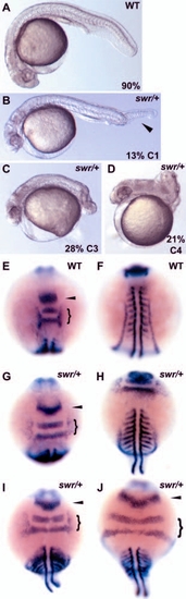
Interaction between sub-dorsalizing tsg1 knockdown and swirl (bmp2b) heterozygotes. All embryos were injected with 16 ng tsg1 MO. At 24 hpf, an injected wild-type embryo was not dorsalized (A), whereas injected swirl (bmp2b) heterozygous siblings were dorsalized. The extent of dorsalization was categorized as follows: (B) weak, class 1 (arrowhead indicates partial loss of the ventral tail fin); (C) moderate, class 3; and (D) strong, class 4. (E-J) Whole-mount in situ hybridization of myod, pax2a and krox20 in 16 ng tsg1 MO1 injected embryos at the eight-somite stage. Injected wild-type embryos were not affected (E,F), whereas heterozygotes displayed a range of dorsalizations (G,H, moderate; I, strong; J, stronger). (F,H) More posterior views of the same embryos shown in E,G, respectively. (J) The somites extend around the circumference, which is typical of a strong, class 4 dorsalization. EXPRESSION / LABELING:
|
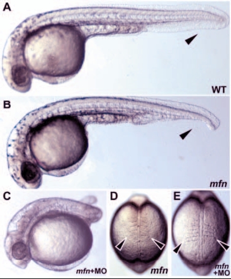
Knockdown of Tsg function enhances dorsalization in mini fin (tolloid) mutants. (A) Wild-type and (B) mfn homozygous mutant embryo at 1 dpf. Arrowheads indicate normal and deficient ventral tail tissue. Injection of 12.5 ng MO5 into mfn/mfn embryos results in a moderate class 3 dorsalization (C). The increase in phenotypic strength is seen at the four-somite stage with the lateral expansion of somites in injected (E) mfn homozygotes (arrowheads) compared with uninjected (D). |
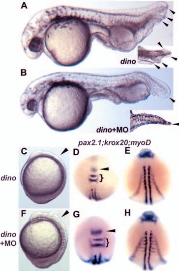
Knockdown of Tsg function partially suppresses the chordino ventralization. (A) Uninjected chordino homozygote; arrowheads indicate multiple ventral fin folds, also seen in a slightly different view at higher magnification (inset). Appearance of multiple fin folds is suppressed by injection of 8 ng MO1 (B, inset shows dorsal view of tail of same embryo). (C-H) Injection of 25 ng MO5 into chordino homozygotes results in a substantial enlargement of dorsally derived tissues. Examination of live embryos at the six-somite stage showed reduced head neural tissue in C and suppression of the reduction in F (arrowheads). (C) Uninjected chordino homozygote; (F) injected chordino homozygote; lateral views, anterior is upwards. In situ analysis at the same stage with pax2.1 (arrowhead), krox20 (bracket) (D,G) and myod (E,H) showed that the somites, the MHB and rhombomeres 3 and 5 are increased in size in the injected (n=14/20) compared with uninjected mutants. (D,E) Uninjected mutants; (G,H) two views of the same injected mutant. In the injected mutant, the anterior neural and somitic mesoderm appears similar to wild type; however, the tail bud is still enlarged, indicating that the dino phenotype is not fully suppressed. EXPRESSION / LABELING:
|
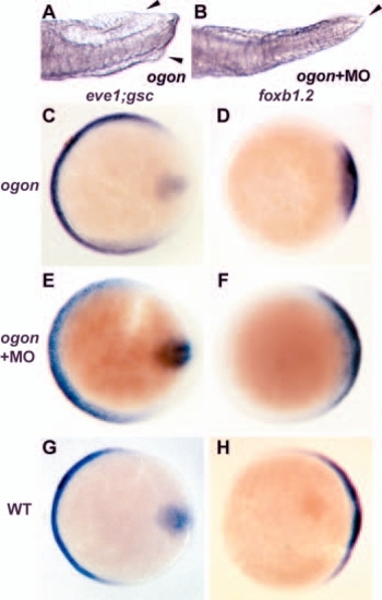
Tsg knockdown partially suppresses the sizzled (ogon) ventralized phenotype. (A) Dorsal view of the tail of an ogon mutant at 24 hpf showing duplication of the ventral fin fold (arrowheads). (B) The fin fold duplication is suppressed by 8 ng MO1. (C-H) Whole-mount in situ hybridization at 80% epiboly (animal pole views, dorsal towards the right) shows ventral marker eve1 expanded in uninjected mutants (C), whereas the dorsal marker foxb1.2 is reduced (D). Expression of these markers in mutants injected with 25 ng MO5 in E (n=16/22) and F (n=15/20) is similar to that seen in wild type (G,H). EXPRESSION / LABELING:
|
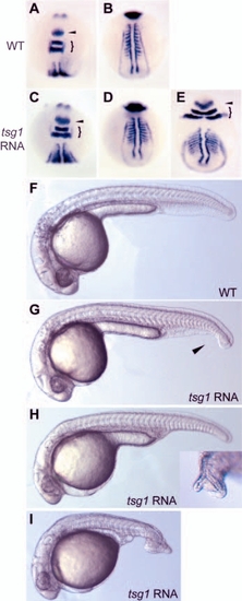
Overexpression of tsg1 RNA causes dorsalization. (A-E) Whole-mount in situ hybridization with pax2.1 (arrowhead), krox20 (brackets) and myod reveals expansion of all three markers in embryos injected with 200 pg tsg1 RNA (C-E) relative to wild type (A,B). (B,D) More posterior views of embryos shown in A and C, respectively. (C,D) Mildly dorsalized and (E) moderately dorsalized embryos. (F-I) At 1 dpf, embryos injected with 100 pg tsg1 RNA show a range of dorsalized phenotypes. (F) Uninjected sibling. (G) Class 1 dorsalization, arrowhead indicates gap in ventral tail fin. (H) Class 2 dorsalization, inset shows duplication in fin fold tissue seen in 50% of class 1 and class 2 embryos. (I) Class 3 phenotype. EXPRESSION / LABELING:
|

Model of Tsg function. |