|
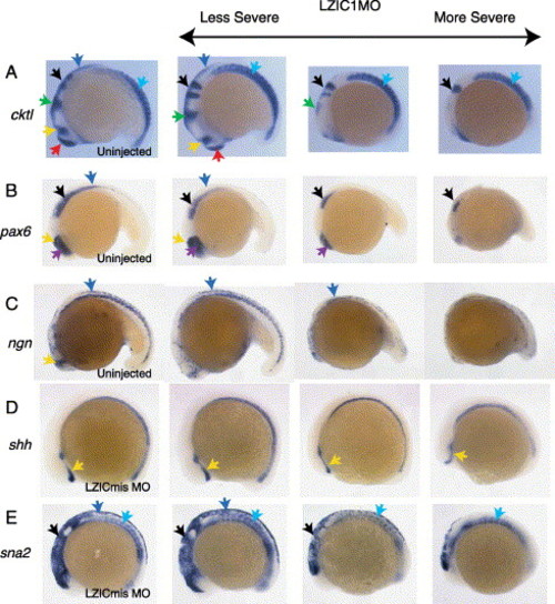
Location of morphant tissue death. Whole mount in situs performed on embryos using a probe cocktail (cktl; A) containing ctsl (anterior ectoderm), opl (forebrain), en2 (midbrain and midbrain/hindbrain boundary), krox20 (rhombomeres three and five), and myoD (somites); pax6 (B); neurogenin (C); shh (D); and sna2 (E). Embryos in columns 2–4 are representative of increasingly severe phenotypes after injection with 3 ng LZIC MO1 at the 1–2 cell stage. Embryos in column 1 are either uninjected or injected with LZIC mismatch control morpholino (LZICmis MO). The CNS is most sensitive to loss of LZIC in the midbrain and spinal column. Telencephalon (red arrows), eye (purple arrows), diencephalon (yellow arrows), midbrain/hindbrain boundary (green arrows), hindbrain (black arrows), dorsal neural tube (blue arrows), somites (turquoise arrows). Lateral views, anterior to left.
|