
- Title
-
Gdf6a is required for the initiation of dorsal-ventral retinal patterning and lens development
- Authors
- French, C.R., Erickson, T., French, D.V., Pilgrim, D.B., and Waskiewicz, A.J.
- Source
- Full text @ Dev. Biol.
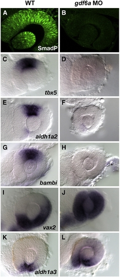
Loss of dorsal signaling and expansion of ventral gene expression in gdf6a morphants. The phosphorylation of Smad 1/5/8 proteins is found in the dorsal retina and lens (A) and is lost in gdf6a morphants (B). The expression of dorsal markers tbx5, aldh1a2, bambi, (C, E, G) are lost in gdf6a morphants (D, F, H). The expression of the ventral specific vax2 (I) and is expanded throughout the retina in gdf6a morphants (J). The expression of the ventral specific aldh1a3 is found also in the ventral retina (K), and is expanded in gdf6a morphants (L). All embryos were staged to 28 hpf. EXPRESSION / LABELING:
|
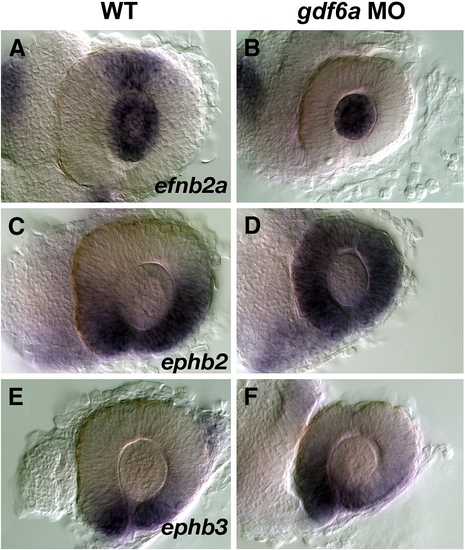
Morpholino inhibition of gdf6a results in aberrant expression of axon guidance molecules. The expression of efnb2a is found in the dorsal retina and lens (A) and is lost in the retina in gdf6a morphants. Expression in the lens is not affected (B). The expression of ephb2 and ephb3 is found in the ventral retina in wildtype animals (C, E), although the domain of epbb3 is more restricted compared to ephb2. The expression of ephb2 is expanded throughout the retina in gdf6a morphants (D), as is the expression of ephb3 (F), albeit to a lesser extent than ephb2. All embryos were staged to 28 hpf. EXPRESSION / LABELING:
|
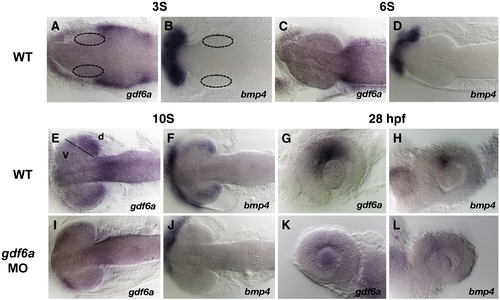
Analysis of Bmp expression in the retina. gdf6a expression is first detected at the 3 somite stage adjacent to the developing eye field (A), while bmp4 expression is limited to the polster (B). The expression domain of gdf6a is reduced by 6 somites, but still located adjacent to the eyecups (C). bmp4 expression is still confined to the polster at 6 somites (D). By 10 somites, both gdf6a and bmp4 expression are found within the presumptive dorsal retina (E, F). Morpholino inhibition of Gdf6a reduces the expression of gdf6a and bmp4 (I, J,). By 28 hpf, both gdf6a and bmp4 are confined to the dorsal retina (G, H), and are down-regulated in gdf6a morphants at 28 hpf (K, L). EXPRESSION / LABELING:
|
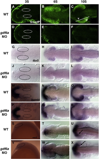
Gdf6a is required for initiation of dorsal–ventral retinal patterning. The phosphorylation of Smad 1/5/8 proteins is not observed in wildtype or morphant embryos at 3 somites (A, D), nor is the expression of the dorsal markers tbx5 and bambi (G, J, M, P). Phosphorylation of Smad proteins is observed by the 6 somite stage (B), and is not detected in gdf6a morphants (E). Phosphorylation becomes more robust by 10 somites in wildtype embryos (C) and is still not detected in gdf6a morphants (F). The expression of tbx5 and bambi is initiated in the retinal tissue at 6 somites (H, N), and is not detected in gdf6a morphants (K, Q). Expression becomes more robust at 10 somites (I, O), and is still not detected in gdf6a morphants (L, R). The expression of vax2 is not detected in either wildtype or morphant animals at 3 somites (S, V). Expression is confined to the forebrain at 6 somites (T) and is not altered in gdf6a morphants (W). Expression of vax2 is detected in the forebrain and presumptive ventral retina in wildtype animals at 10 somites (U), and is mildly expanded in gdf6a morphants (X). Dotted circles delineate the optic cup. Black arrows point to the expanded expression of vax2 in gdf6a morphants. EXPRESSION / LABELING:
|
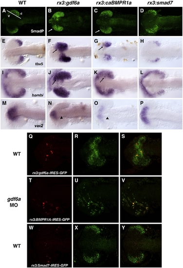
Over-expression of Gdf6a signaling components induces dorsal identity and represses ventral identity. The phosphorylation of Smad 1/5/8 proteins is induced in the presumptive ventral retina when either gdf6a or BMPR1a is expressed using the Medaka rx3 promoter (B, C). This effect is cell non-autonomous with respect to gdf6a, as phosphorylation is induced in cells surrounding those that express rx3:gdf6a-IRES-GFP (Q–S). Expression of caBMPR1a exerts its effects in a cell autonomous fashion as phosphorylation is detected specifically in cells that express rx3:caBMPR1a-IRES-GFP (T–V). The expression of tbx5 (E, F) and bambi (I, J) are also induced ectopically in the retina under both conditions. The expression of vax2 is found in the retina just proximal to the forebrain in wildtype embryos (M), and this expression is lost in embryos expressing either gdf6a (N) or BMPR1a (O). Expression of the inhibitory smad7 via the rx3 promoter inhibits phosphorylation of Smad 1/5/8 proteins (D), as well as the expression of tbx5 (H) and bambi (L). Smad7 works in a cell autonomous fashion as Smad phosphorylation is specifically inhibited in cells expressing rx3:smad7-IRES-GFP (W–Y). Expression of smad7 has no effect on the retinal expression of vax2 (P). All embryos were staged to 10 somites. White arrows indicate ectopic Smad 1/5/8 phosphorylation in the presumptive ventral retina, while black arrows indicate ectopic expression of dorsal genes in the presumptive ventral retina. Arrowheads point to the reduction of vax2 expression in the presumptive ventral retina in embryos over-expressing gdf6a or caBMPR1a. EXPRESSION / LABELING:
|

Morpholino inhibition of Tbx5 affects dorsal, but not ventral gene expression. The retinal expression of efnb2a is lost in tbx5 morphants, while the lens-specific expression remains unchanged (A, B). The expression aldh1a2 is lost (C, D), while the expression of bambi is highly down-regulated (E, F). In the ventral retina, vax2 (G and H), ephb2 (I and J), and aldh1a3 (K, L) are not altered in tbx5 morphants. All embryos were staged to 28 hpf. EXPRESSION / LABELING:
|
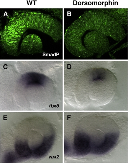
Dorsomorphin inhibits Smad phosphorylation and tbx5 expression. The nuclear localized Smad 1/5/8 is reduced upon incubation in 100 μM Dorsomorphin (A, B). The expression of tbx5 is reduced in these embryos (C, D). The dorsal limit of vax2 expression is slightly raised upon incubation in Dorsomorphin (E, F). All embryos were staged to 28 hpf. EXPRESSION / LABELING:
|
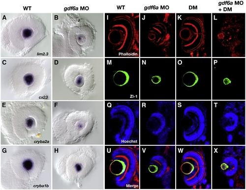
Lens-specific gene expression is deregulated in gdf6a morphants. The expression of lim2.3 is down-regulated at 28 hpf in gdf6a morphant embryos (A, B), as is the expression of cx23 (C, D) and crystallin ba2a (E, D). The expression of the related crystallin ba1b is also down-regulated (G, H). At 4 dpf, there is little difference in the organization of F-actin in gdf6a morphants, as no changes in phalloidin staining are observed (I, J). There is also no change in nuclear staining, indicating no residual nuclear retention in gdf6a morphants (Q, R). There is however a slightly higher level of Zl-1 staining in the morphant lens, which identifies differentiating lens fibers (M, N). Incubation in Dorsomorphin has no effect of lens development (K, O, S, W), but when combined with gdf6a morpholino inhibition, ectopic phalloidin and Zl-1 is observed in the lens (L, P), and there is evidence of nuclear retention (T). A merged image of all staining is shown in panels U–X. |
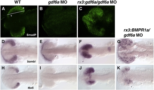
Expression of gdf6a under the transcriptional control of the medaka rx3 promoter can rescue Smad 1/5/8 phosphorylation in gdf6a morphants (compare C with B), to near wildtype levels (A). Similarly, the expression of bambi and tbx5 is rescued by expressing either rx:gdf6a (F and J) or rx3:BMPR1a (G and K). EXPRESSION / LABELING:
|
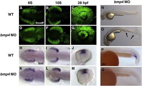
Morpholino inhibition of bmp4 has no effect on eye patterning. Phosphorylation of Smad 1/5/8 proteins is not affected at any stage tested, including 6 somites (A, D), 10 somites (B, F), or 28 hpf (C, G). Similarly, the expression of tbx5 is normal at 6 somites (H, K) 10 somites (I, L) and 28 hpf (J, M). Morphological phenotypes of bmp4 morphants at 2 dpf (compare O with N) showing a C2 dorsalized phenotype (Mullins et al., 1996) involving loss of the ventral tail fin and ventral vein. Expression of a Bmp4 target gene, flk1 24 hpf, is down-regulated in bmp4 morphants (compare Q with P). Both phenotypes are observed in a previously published bmp4 null mutant (Stickney et al., 2007). Arrows denote the extent of ventral tissue loss. EXPRESSION / LABELING:
|

Unillustrated author statements |
Reprinted from Developmental Biology, 333(1), French, C.R., Erickson, T., French, D.V., Pilgrim, D.B., and Waskiewicz, A.J., Gdf6a is required for the initiation of dorsal-ventral retinal patterning and lens development, 37-47, Copyright (2009) with permission from Elsevier. Full text @ Dev. Biol.