- Title
-
Lbx2 regulates formation of myofibrils
- Authors
- Ochi, H., and Westerfield, M.
- Source
- Full text @ BMC Dev. Biol.
|
Muscle precursors express lbx1b and lbx2. A-L: Muscle precursors transiently express lbx2. A-G, L: Expression of lbx2 at 70%-epiboly (A), 90%-epiboly (B), bud (C), segmentation (D), 24 hpf (E, L), 48 hfp (F, G). Whole-mounts. H-K: lbx2 (blue) and ntl (red) expression at 70%-epiboly (H), 90%-epiboly (I), bud (J), and segmentation (K). Flat-mounts. L: Transverse section, 24 hpf embryo, F59 (green) and lbx2 mRNA (blue). (A, H) lbx2 mRNA appears at 70%-epiboly adjacent to ntl expressing cells in blastoderm margin. lbx2 expression in paraxial mesoderm by end of gastrulation (B-C, I,). lbx2 expression later restricted to adaxial cells (J-K, brackets). (L) Subset of fast muscle cells expresses lbx2 in epaxial (black arrow) and hypaxial (black arrowhead) domains. F59 and lbx2 labeling shows differentiated slow muscle cells lose lbx2 expression (L, white arrowhead). (F) lbx2 expression in trunk disappears by 48 hpf (F, bracket). lbx2 expression in fin primordia (F-G, black arrowheads), hindbrain (F-G, white arrowhead), and hyoid (F, white arrow). M: Diagram of zebrafish muscle. Adaxial cells (K, bracket) migrate superficially and differentiate into slow muscle fibers (green). Then, fast (magenta) and medial fast fibers (red) differentiate. N-R: 5-somite (N), 18-somite (O), 24 hpf (P-Q) and 48 hpf (R). (Q) Higher magnification of P. lbx1b mRNA appears at 5-somite stage in neural tube (N), along rostral-caudal axis (O, arrow), then lateral to somites (P-Q), and later in fin (R). (A-D, N-O) Dorsal views, rostral towards the top; (E-F, P) lateral views, rostral toward the left, dorsal toward the top; (H-K) rostral toward the top; (L) dorsal toward the top; (G, R) dorsal views, rostral toward the left. Scale bars: (A-D, N-O, Q) 200 μm, (E-G, P, R) 100 μm, (H-L) 50 μm. EXPRESSION / LABELING:
|
|
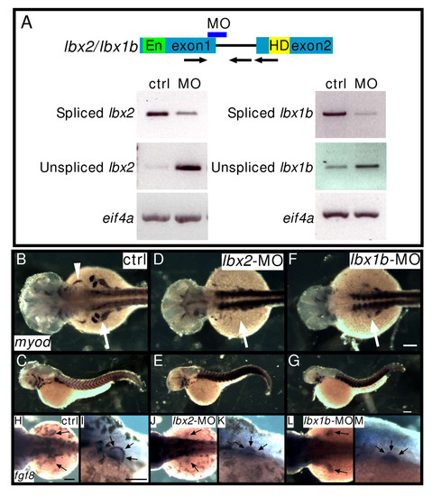
Lbx2 and Lbx1b function in hypaxial muscle development. A: Splice donor MOs against lbx2 or lbx1b inhibit correct splicing of lbx2 or lbx1b, respectively. RT-PCR was performed using bud stage (lbx2) or segmentation stage (lbx1b) embryos. Spliced bands and unspliced bands in the lbx-splice donor-MO lane indicate aberrantly spliced message increases and correctly spliced message decreases. Arrows indicate primers. En: Engrailed domain, HD: homeodomain. Arrows indicate specific primers for amplification of correctly spliced or unspliced lbx genes. B-G: Expression of myod in control (ctrl) embryos (B, C), lbx2-MO injected embryos (D, E). lbx1b-MO injected embryos (F, G). The white arrowhead indicates the sternohyoideus primordium (B) and the white arrows indicate fin muscle precursors. myod expression in fin bud is suppressed by lbx2-MO or lbx1b-MO (B: 100%, n = 36; D: 12%, n = 56; F: 16%, n = 24). H-M: Expression of fgf8 in ectodermal cells of the fin bud. Control embryos (H, I, 100%, n = 12), lbx2-MO injected embryos (J-K, 100%, n = 12), lbx1b-MO injected embryos (L-M, 100%, n = 15). (B, D, F, H, J, L) Dorsal views, rostral towards the left, (C, E, G. I, K, M) lateral views, rostral toward the left, dorsal toward the top. Scale bar: (B-M) 100 μm. EXPRESSION / LABELING:
|
|
Lbx2 is not required for the induction of myogenesis. A-D: Suppression of Lbx2 activity does not affect myod or myf5 expression. The adaxial cells (precursors of slow muscles and muscle pioneers) express myod (A, 100%, n = 20). Embryos injected with lbx2-MO express myod at normal levels (B, 100%, n = 20). The paraxial mesoderm expresses myf5 in control embryos (C, 100%, n = 17) and lbx2-MO injected embryos (D, 100%, n = 12). E-F: Adaxial cell morphology at 3-somite stage in control (E) or lbx2-MO injected (F) embryos. Nomarski images of dorsal views. The brackets indicate the width of the adaxial cell row. The normally cuboidal adaxial cells are aberrantly shaped in the lbx2-MO injected embryo. Arrows indicate individual adaxial cells. G-H: Several myod expressing cells fail to incorporate properly into the adaxial cell monolayer after Lbx2 knockdown. (A-H) Whole-mount embryos, dorsal views, rostral toward the top. Scale bar: (A-D) 200 μm (E, F) 25 μm, (G, H) 50 μm. EXPRESSION / LABELING:
PHENOTYPE:
|
|
Knockdown of Lbx2 activity results in malformation of slow muscle fibers. A-D: Control embryos. E-H: lbx2-MO injected embryos. I-L: lbx2 mRNA injected embryos. (A-B, E-F, I-J) Embryos labeled with the slow muscle marker, F59 (red). (B, F, J) Higher magnification. Rostral-caudal extension of slow fibers is severely affected by lbx2 knockdown (B, F, arrows). M: Statistical analyses of mean filament rostral-caudal length (arrows) and dorsoventral thickness (B, F, arrowheads). Analysis by ANOVA demonstrates significant differences in length (P < 0.01) and thickness (P < 0.05). (C, G, K, N) Embryos labeled with Prox1 (green, nuclear slow muscle and muscle pioneer marker), and 4D9 (red, Eng, muscle pioneer marker,). Green indicates slow muscle cells and yellow shows muscle pioneers (MP). (ctrl: n = 10, lbx2-MO: n = 6, lbx2-mRNA: n = 4). No apparent differences can be detected between controls and embryos injected with lbx2-MO. N: Analysis by ANOVA demonstrates no significant differences. The data represent the average ± s.e.m. (D, H, L) Transverse sections of a 24 hpf embryo labeled with the slow muscle marker, F59 (green) and Hoechst to mark nuclei. Slow muscle cells migrate properly to the superficial layer. (A-C, E-G, I-K) Lateral views, rostral toward the left, dorsal toward the top; (D, H, L) dorsal toward the top. Scale bar: (A, C-E, G-I, K-L) 50 μm; (B, F, J) 25 μm. |
|
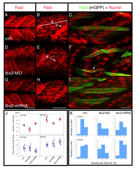
Fast muscle fibers are malformed in the absence of Lbx2. A-C: Control embryos. D-F: lbx2-MO injected embryos. G-I: lbx2 mRNA injected embryos. (A, B, D, E, G, H, J) Embryos labeled with the fast muscle marker, EB165 (red). (B, E, H) Higher magnification views. Rostral caudal extension of fast fibers is severely affected (B, E, arrow). J: Statistical analyses of mean filament rostral-caudal length (B, E, arrows) or dorsal ventral thickness (B, E, arrowhead). Analysis by ANOVA demonstrates significant differences in length (P < 0.01). (C, F, I) Embryos were mosaically labeled with membrane localized GFP (mGFP) by injection of DNA and labeled for nuclei (red, propidium iodide). mGFP expressing cells in the fast muscle domain at the level of somite 10 were randomly selected and the number of their nuclei counted. K: Fast muscle cells are multinucleate by 48 hpf in both control and lbx2 mRNA injected embryos, whereas unfused fast muscle cells are observed in lbx2-MO injected embryo (F, arrow). Analysis demonstrates significant differences among the three groups at 48 hpf (Kruskal-Wallis test, p = 0.0121). No apparent differences can be detected between controls and embryos injected with lbx2-MO at 24 hpf (Kruskal-Wallis test, p = 0.1509). (A-I) Lateral views, rostral toward the left, dorsal toward the top. Scale bars: (A, B, D, E, G, H) 50 μm, (C, F, I) 20 μm. |
|
Expression of thin and thick myofilament genes is downregulated by interference with Lbx2 activity. A-J: Analysis of myofilament gene expression. Expression of acta1 (A), tnnt1 (B), tpma (C), tnnt3b (D), tnnc (E), myhz1 (F), mybpc1 (G), smyhc (H), myhz2 (I), mylz2 (J) in control, lbx2-MO injected, lbx2 mRNA injected, and Engrailed suppresser domain fused lbx2 (EnR-lbx2) mRNA injected embryos. Expression of acta1 (A, ctrl: 28/28, lbx2-MO: 0/33, lbx2-mRNA: 9/10, EnR-lbx2 mRNA: 8/8, numerator indicates number of embryos with normal expression, and denominator indicates number of examined embryos), tnnt1 (B, ctrl: 19/19, lbx2-MO: 7/22, lbx2-mRNA: 12/12, EnR-lbx2 mRNA: 17/17), tpma (C, ctrl: 16/16, lbx2-MO: 2/20, lbx2-mRNA: 13/14, EnR-lbx2 mRNA: 10/11) and tnnt3b (D, ctrl: 13/13, lbx2-MO: 0/7, lbx2-mRNA: 7/7, EnR-lbx2 mRNA: 4/4) is reduced by lbx2-MO, but not tnnc (E, ctrl: 17/17, lbx2-MO: 18/22, lbx2-mRNA: 10/10, EnR-lbx2 mRNA: 16/16). Expression of myhz1 (F, ctrl: 25/25, lbx2-MO: 3/16, lbx2-mRNA: 7/8, EnR-lbx2 mRNA: 8/8), mybpc1 (G, ctrl: 15/15, lbx2-MO: 10/20, lbx2-mRNA: 13/14, EnR-lbx2 mRNA: 5/8) and myhz2 (I, ctrl: 17/17, lbx2-MO: 0/5, lbx2-mRNA: 9/9, EnR-lbx2 mRNA: 8/8) is reduced by lbx2-MO, but smyhc (H, ctrl: 36/36, lbx2-MO: 24/29, lbx2-mRNA: 8/8, EnR-lbx2 mRNA: 12/12) and mylz2 are unaltered (J, ctrl: 15/15, lbx2-MO: 14/15, lbx2-mRNA: 10/10, EnR-lbx2 mRNA: 7/7). No obvious difference can be detected between embryos injected with lbx2 mRNA and embryos injected with EnR-lbx2 mRNA. (A-J) Whole mounts, lateral views, rostral toward the left, dorsal toward the top. Scale bar: 100 μm. EXPRESSION / LABELING:
|
|
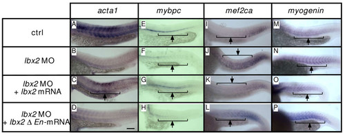
The Engrailed domain of Lbx2 is required for myofilament gene expression. A-H: Expression of acta1 (A-D) and mybpc1 (E-H). Embryos express acta1at 24 hpf (A: 93% with normal expression, n = 15). Injection of lbx2-MO results in decreased acta1 expression (B: 20% with normal expression, n = 20), whereas acta1 expression partially recovers in embryos injected with lbx2-MO + lbx2 mRNA (C: 42% with normal expression, n = 50). In contrast, embryos injected with lbx2-MO + lbx Δ En mRNA fail to express acta1 (D: 14% with normal expression, n = 37). mybpc1 (E: 94%, n = 18; F: 43%, n = 23; G: 81%, n = 31; H: 42%, n = 38). I-P: Expression of mef2ca (I-L) and myogenin (M-P). Embryos express mef2ca at 24 hpf (I: n = 11). Injection of lbx2-MO results in increased mef2ca expression (J: 72% with increased expression, n = 11), whereas mef2ca expression partially recovers in embryos injected with lbx2-MO + lbx2 mRNA (K: 21% with increased expression, n = 19). In contrast, embryos injected with lbx2-MO + lbx Δ En mRNA fail to express mef2ca (L: 61% with increased expression, n = 21). myogenin (M: n = 7, N: 78% with increased expression, n = 9, O: 23%, n = 13, P: 67%, n = 14). (A-P) Whole mounts, lateral views, rostral toward the left, dorsal toward the top. Scale bar: 100 μm. EXPRESSION / LABELING:
|
|
Cells with impaired motility due to Lbx2 knockdown still express myod.lbx2-MO and rhodamine (red) injected cells were transplanted into the margin, 75 degrees ventral to the shield, at shield stage. When control cells are transplanted into control embryos, the cells become distributed along the rostral caudal axis (A) and transplanted cells adjacent to the notochord express myod (A inset, arrow, ctrl > ctrl: n = 4). In contrast, transplanted lbx2-MO injected cells stay clumped and do not distribute along the rostral caudal axis, although cells adjacent to the notochord express myod normally (B inset, arrow, lbx2-MO > ctrl: n = 4/4). This effect on migration appears to be cell-autonomous because control cells transplanted into lbx1b-MO injected embryos behave normally (C, ctrl > lbx2-MO: n = 2). lbx2-MO + lbx2-mRNA injected cells become distributed along the rostral caudal axis (D, n = 13/15 rescued). Arrows indicate myod expressing transplanted cells. N: notochord. (A-D) Whole-mount embryos, dorsal views, rostral toward the top. Scale bar: 200 μm. |
|
Absence of Lbx2 activity results in malformation of slow and fast muscle fibers in lbx2 translation MO injected embryos. (A, B, E, F) Control embryos. (C, D, G, H) lbx2 translation MO injected embryos. Embryos labeled with the slow muscle marker, F59 (A-D) and the fast muscle marker, EB165 (E-H). (A-H) Whole mounts, lateral views, rostral toward the left, dorsal toward the top. Scale bars: (A, C, E, G) 50 μm, (B, D, F, H) 20 μm. |
|
Lbx2 regulates slow and fast muscle fiber formation. (A-B) Embryos labeled with α-actin (red) and F59, slow MyHC (green). Dotted lines indicate somite borders. 24 hpf embryos. (C-H) Embryos labeled with the slow muscle marker, F59 (C, D, E) or the fast muscle marker EB165 (F, G, H). 48 hpf embryos. (A-H) Whole mounts, lateral views, rostral toward the left, dorsal toward the top Scale bar: (A, B) 12.5 μm; (C-H) 50 μm. |
|
myogenin, mef2ca and pax7 are downstream targets of Lbx2. (A-C) Expression of myogenin (A), mef2ca (B) and pax7 (C) in control (ctrl), lbx2-MO injected embryos and lbx2 mRNA injected embryos. The expression of myogenin (A) and mef2ca (B) are suppressed by overexpression of lbx2 mRNA. In contrast, expression of pax7 is suppressed by lbx2-MO (C). myogenin (A, ctrl: 14/14 with normal expression, lbx2-MO: 13/15, lbx2-mRNA: 0/18), mef2ca (B, ctrl: 11/11, lbx2-MO: 4/5, lbx2-mRNA: 0/8), pax7 (D, ctrl: 7/7, lbx2-MO: 0/14, lbx2-mRNA: 13/13). (A-F) Whole mounts, lateral views, rostral toward the left, dorsal toward the top. Scale bar: 100 μm. |


