
Fig. 3
- ID
- ZDB-IMAGE-090320-41
- Genes
- Publication
- Ochi et al., 2009 - Lbx2 regulates formation of myofibrils
- All Figures
- Figures for Ochi et al., 2009
|
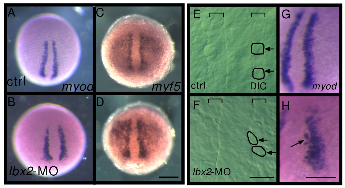
Fig. 3 Lbx2 is not required for the induction of myogenesis. A-D: Suppression of Lbx2 activity does not affect myod or myf5 expression. The adaxial cells (precursors of slow muscles and muscle pioneers) express myod (A, 100%, n = 20). Embryos injected with lbx2-MO express myod at normal levels (B, 100%, n = 20). The paraxial mesoderm expresses myf5 in control embryos (C, 100%, n = 17) and lbx2-MO injected embryos (D, 100%, n = 12). E-F: Adaxial cell morphology at 3-somite stage in control (E) or lbx2-MO injected (F) embryos. Nomarski images of dorsal views. The brackets indicate the width of the adaxial cell row. The normally cuboidal adaxial cells are aberrantly shaped in the lbx2-MO injected embryo. Arrows indicate individual adaxial cells. G-H: Several myod expressing cells fail to incorporate properly into the adaxial cell monolayer after Lbx2 knockdown. (A-H) Whole-mount embryos, dorsal views, rostral toward the top. Scale bar: (A-D) 200 μm (E, F) 25 μm, (G, H) 50 μm.