
|
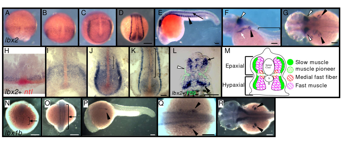
Fig. 1 Muscle precursors express lbx1b and lbx2. A-L: Muscle precursors transiently express lbx2. A-G, L: Expression of lbx2 at 70%-epiboly (A), 90%-epiboly (B), bud (C), segmentation (D), 24 hpf (E, L), 48 hfp (F, G). Whole-mounts. H-K: lbx2 (blue) and ntl (red) expression at 70%-epiboly (H), 90%-epiboly (I), bud (J), and segmentation (K). Flat-mounts. L: Transverse section, 24 hpf embryo, F59 (green) and lbx2 mRNA (blue). (A, H) lbx2 mRNA appears at 70%-epiboly adjacent to ntl expressing cells in blastoderm margin. lbx2 expression in paraxial mesoderm by end of gastrulation (B-C, I,). lbx2 expression later restricted to adaxial cells (J-K, brackets). (L) Subset of fast muscle cells expresses lbx2 in epaxial (black arrow) and hypaxial (black arrowhead) domains. F59 and lbx2 labeling shows differentiated slow muscle cells lose lbx2 expression (L, white arrowhead). (F) lbx2 expression in trunk disappears by 48 hpf (F, bracket). lbx2 expression in fin primordia (F-G, black arrowheads), hindbrain (F-G, white arrowhead), and hyoid (F, white arrow). M: Diagram of zebrafish muscle. Adaxial cells (K, bracket) migrate superficially and differentiate into slow muscle fibers (green). Then, fast (magenta) and medial fast fibers (red) differentiate. N-R: 5-somite (N), 18-somite (O), 24 hpf (P-Q) and 48 hpf (R). (Q) Higher magnification of P. lbx1b mRNA appears at 5-somite stage in neural tube (N), along rostral-caudal axis (O, arrow), then lateral to somites (P-Q), and later in fin (R). (A-D, N-O) Dorsal views, rostral towards the top; (E-F, P) lateral views, rostral toward the left, dorsal toward the top; (H-K) rostral toward the top; (L) dorsal toward the top; (G, R) dorsal views, rostral toward the left. Scale bars: (A-D, N-O, Q) 200 μm, (E-G, P, R) 100 μm, (H-L) 50 μm.