- Title
-
Zebrafish foxd3 is selectively required for neural crest specification, migration and survival
- Authors
- Stewart, R.A., Arduini, B.L., Berghmans, S., George, R.E., Kanki, J.P., Henion, P.D., and Look, A.T.
- Source
- Full text @ Dev. Biol.
|
sym1 mutants have defects in trunk and vagal neural crest-derived peripheral neurons, cranial ganglion neurons and glia. Lateral views of wild-type (A, B) and sym1 mutant (E, F) embryos showing tyrosine hydroxylase (th) expression at 5 dpf (A, E) and 48 hpf (B, F). There is a dramatic reduction in the number of th-positive cells in the region of the developing sympathetic cervical complex (arrowheads) in sym1 mutants (0–2 cells at 2 dpf, 0–10 cells at 5 dpf, n = 10), compared to their wild-type siblings (10–15 cells at 2 dpf, 40–50 cells at 5 dpf, n = 10). High magnification ventral views of wild-type (C, D) and sym1 mutant (G, H) embryos, showing that the loss of sympathetic neurons in sym1 mutants is reflected by a severely reduced number of sympathoblasts based on zash1a expression at 40 hpf (C, G) and phox2b expression at 48 hpf (D, H). Arrows in C, D, G, H indicate migrating sympathoblasts. Lateral views of wild-type (I–L) and sym1 mutant (M–P) embryos. Black arrows indicate position of developing DRG (I) and enteric neurons (J) in 3 dpf wild-type embryos, which are largely absent in sym1 mutants (M, N). The cranial ganglion neurons at 3 dpf are reduced in size in sym1 mutants by approximately 30% (K compared to O). White arrows indicate the trigeminal (tg) and posterior lateral line (pllg) ganglia complex. Black arrows indicate epibranchial ganglia (eb). (L, P) foxd3 is expressed in developing glia associated with the posterior lateral line ganglia (arrow) and axon tract (arrowhead) in wild-type embryos at 48 hpf (L), but is severely reduced or absent in sym1 mutant embryos (P). Abbreviations: absence of expressing cells (*), amacrine cells (am), axon tract (at), dopaminergic neurons (dp), hindbrain (hb), neural crest-derived enteric and sympathetic precursors (NC), sympathetic neurons (sym). |
|
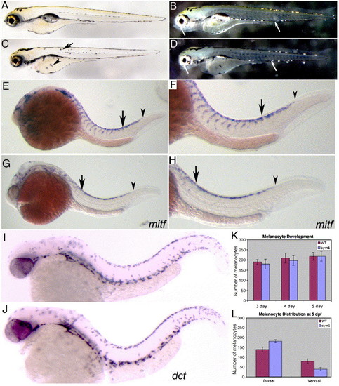
Development of neural crest-derived chromatophores in sym1 mutants. Lateral views of living wild-type (A, B) and sym1 mutant (C, D) embryos at 3 dpf. Melanophores (black) are evident in both wild-type (A) and sym1 (C) mutant embryos, although sym1 mutants have more dorsally-displaced melanocytes on the trunk (arrow) and yolk (arrowhead; see also panel L). Incident light reveals silver iridiphores are reduced in some sym1 mutants (D). Lateral views of developing melanoblasts expressing mitf in wild-type (E, F) and sym1 mutant (G, H) embryos. F and H are higher magnification views of the trunk region in E and G. Arrowheads in E–H indicate the posterior extent of mitf staining along the trunk in wild-type and sym1 mutants, while the arrows in E–H show the posterior extent of dorsal–ventral neural crest migration, both of which are delayed in sym1 mutants (compare E, F with G, H). Expression of dct at 36 hpf in wild-type (I) and sym1 mutant (J) embryos. The expression pattern and numbers of dct-positive cells are indistinguishable between wild-type and sym1 mutants. Quantitation of melanophore numbers (K) and distribution (L) in wild-type (red) and sym1 mutants (blue). Abbreviations: mitf (microphthalmia-associated transcription factor), dct (dopachrome tautomerase). |
|
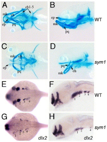
Abnormal pharyngeal arch development in sym1 mutants. Wild-type (A, B) and sym1 mutant (C, D) embryos at 5 dpf, stained with Alcian blue to reveal craniofacial cartilages. Ventral (A, C) and lateral (B, D) views show that the 1st (mk) and 2nd (ch) arch derivatives are present but abnormal in sym1 mutants, whereas 3rd arch-derived cartilages (cb1–5) are missing altogether. dlx2 expression in migrating neural crest pharyngeal arch precursors, shown in dorsal views at 24 hpf in wild-type (E) and sym1 mutant (G) embryos, and in lateral views of 32 hpf wild-type (F) and sym1 mutant (H) embryos. sym1 mutants have reduced dlx2 expression in migrating neural crest cells that generate the 1st and 2nd arch derivatives (labeled 1 and 2), and more severely reduced dlx2 expression in neural crest cells that generate the pharyngeal cartilages (labeled 3 and 4; G, H). Abbreviations: Meckel's cartilage (mk), palatoquadrate (pq), ceratohyal arch (ch), ethmoid plate (ep), and ceratobranchial (cb) arches 1–5. |
|
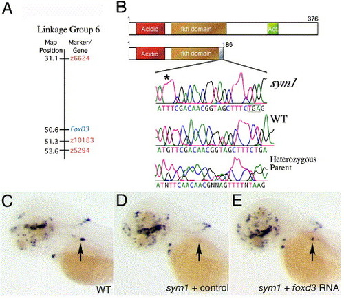
sym1 is an inactivating mutation in foxd3. (A) Linkage of the foxd3 gene to the CA repeat markers on zebrafish chromosome 6. The positions of markers z10183, z6624 and z5294, are highlighted in orange (source http://zfin.org), while foxd3 is highlighted in blue. The z10183 marker was mapped to within 1 cM of the foxd3 gene. (B) Schematic diagrams of the wild-type and mutant Foxd3 proteins. In sym1, a guanine (537) deletion results in a frame shift leading to a premature stop codon and disruption of the encoded DNA-binding domain. An acidic domain is highlighted in orange at the N-terminus, and the C-terminus transcriptional effector domain is shown in green. Chromatogram traces below the schematic illustrate the nucleotide change (asterisk) affecting the foxd3-coding region. (C–E) Injection of foxd3 RNA rescues the sympathetic th-expression pattern in sym1 mutant embryos. Dorso-lateral views at 4 dpf of wild-type (C) and sym1 mutant embryos (D, E) labeled with the th riboprobe. Wild-type siblings contain 10–40 th-expressing cells in the cervical complex at this stage (C), whereas the sym1 mutant embryos injected with control RNA have 0–2 th-expressing cells (D). Injection of full-length foxd3 RNA into sym1 mutant embryos results in rescue (E), defined as >10 th-expressing sympathetic neurons in the region of the cervical complex. Arrows indicate position of the cervical complex. EXPRESSION / LABELING:
|
|
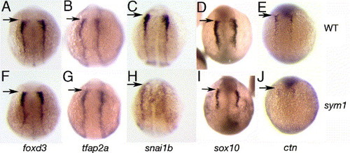
Molecular defects in neural crest specification in sym1 mutants. Dorsal views of wild-type (A–E) and sym1 mutant (F–J) embryos. Neural plate border expression of foxd3 (A, F), tfap2a (B, G), snai1b (C, H), sox10 (D, I) and ctn (ctn E, J) in 3-somite-stage embryos. Decreased expression of sox10, snai1b and ctn, but not foxd3 and tfap2a is evident in sym1 mutants. Arrows indicate position of midbrain–hindbrain boundary. EXPRESSION / LABELING:
|
|
Gene expression abnormalities in premigratory and early migrating neural crest cells in sym1 mutants. Lateral, cranial and trunk views of wild-type (A–C, G–I, M–O) and sym1 (D–F, J–L, P–R) embryos, showing expression of tfap2a (A–F), sox10 (G–L), and snai1b (M–R) at the 10-to 12-somite stage. tfap2a expression begins to decrease in sym1 mutants at this stage, particularly in the trunk (compare A, C with D, F). The expression of sox10 in sym1 mutants (J–L) and snai1b (P–R) is reduced in both cranial and trunk neural crest. The reduction in sox10 expression is specific to the neural crest because sox10 expression in the developing otic placode (arrowheads) is normal in wild-type and sym1 mutants (compare H with K). Arrows indicate the position of the trunk neural crest. EXPRESSION / LABELING:
|
|
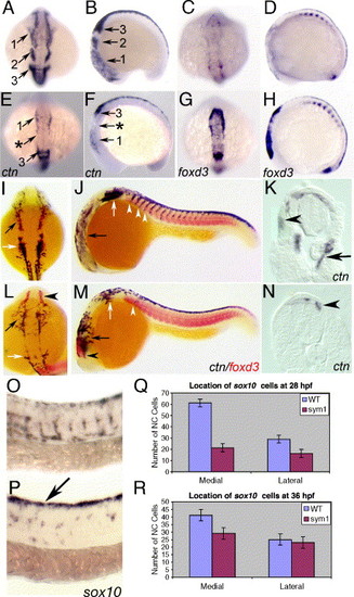
Abnormal cell migration in sym1 mutant embryos. Wild-type (A–D) and sym1 mutant (E–H) embryos at the 12-somite stage, showing dorsal (A, E, C, G) and lateral (B, F, D, H) views. ctn expression reveals migration of the three cranial neural crest streams in wild-type embryos (A–B, numbered arrows). In sym1 embryos, ctn expression in cranial neural crest is severely reduced in the 1st neural crest stream (E, F) and almost absent in the 2nd stream (arrow with asterisk in E, F). ctn-positive cells of the 3rd stream are present in sym1 mutants, but only a few cells migrate ventrally (compare F with B). At the same stage, foxd3 expression is normally rapidly downregulated in wild-type embryos upon migration (C–D), but persists in sym1 mutants (G–H), showing that the cranial premigratory crest is present, but does not migrate. Wild-type (I–J) and sym1 mutant (L–M) embryos double labeled with ctn (black) and foxd3 (red) at the 26-somite stage, showing dorsal (I, L) and lateral (J, M) views. At this stage, ctn is expressed throughout the head in deep ventral positions of wild-type embryos (I, J, black arrow); however, in sym1 mutants, ctn-positive cells fail to migrate ventrally, and spread out more laterally (L, M, black arrow). The expression of ctn is also strongly reduced in the region of the hindbrain posterior to the otic vesicle in sym1 mutants (L–M, white arrows), while foxd3 expression remains high in the anterior-most neural crest cells (L, M, black arrowhead). Numerous, ctn-expressing trunk neural crest cells are seen migrating in wild-type embryos at the 26-somite stage (J; white arrowhead), but few ctn-positive cells are seen in sym1 embryos (M; white arrowhead). Transverse sections at the anterior trunk level of 24 hpf wild-type (K) and sym1 mutant (N) embryos, showing ctn expression in both dorsal-laterally (K, arrowhead) and medial (K, arrow) migrating neural crest cells in wild-type embryos. In sym1 mutants, migration of ctn-expressing cells are delayed, and found only in dorsal positions at this stage (N, arrowheads). Lateral views of sox10-expressing neural crest cells in wild-type (O) and sym1 (P) embryos at 24 hpf. In wild-type embryos, robust sox10 expression is evident in migrating neural crest cells; however, in sym1 mutants most of the sox10-expressing cells are still on the dorsal neural tube (P, arrow). Quantitation of sox10-positive cells located along each migration pathway at 28 hpf (Q) and 36 hpf (R) in wild-type (blue) and sym1 mutants (red), shows that migration along the medial pathway is more severely affected in sym1 mutants. EXPRESSION / LABELING:
|
|
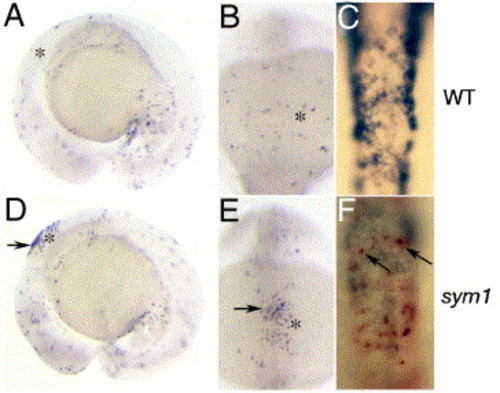
Neural crest cell death in sym1 mutants. Lateral (A, D) and dorsal (B, C, E, F) views of TUNEL labeled 15-somite stage wild-type (A–C) and sym1 mutant (D–F) embryos shows an increase in dorsal TUNEL-positive cells in the hindbrain region in sym1 mutants (arrows in D, E). Asterisks indicate the position of the developing otic placode. (C, F) Double labeling analysis with ctn (dark blue) and TUNEL (red) shows cell death in some neural crest cells that still express ctn in sym1 mutants at this stage (arrows). |
|
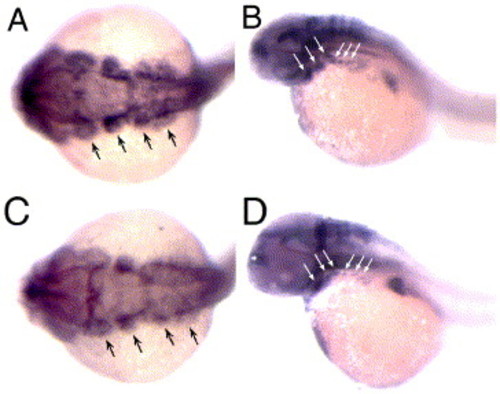
Abnormal pharyngeal arch development in sym1 mutants. Wild-type (A, B) and sym1 mutant (C, D) embryos. (A, C) Dorsal views of sox9a expression in 24 hpf embryos. Black arrows indicate cranial arch streams of neural crest cells. (B, D) Lateral views of sox9a expression in 48 hpf wild-type (B) and sym1 mutant (D) embryos. White arrows indicate arch neural crest streams. At both 24 and 48 hpf, sox9a arch expression is reduced in sym1 mutants, particularly in posterior (branchial) arches. EXPRESSION / LABELING:
|
|
Normal somite and floorplate development in wild-type (A–C) and sym1 mutant (D–F) embryos. (A, D) Dorsal views showing normal myoD expression in axial and paraxial mesoderm in 18-somite stage wild-type (A) and sym1 mutant (D) embryos. (B, C, E, F) Lateral views show normal floor plate expression of twhh (B, E, arrows) in 20-somite stage embryos and col2a1 (C, F, arrows) in 24 hpf embryos. Abbreviations: myoD (myogenic differentiation D), twhh (tiggy winkle hedgehog), col2a1 (collagen type II, alpha-1a). EXPRESSION / LABELING:
|
|
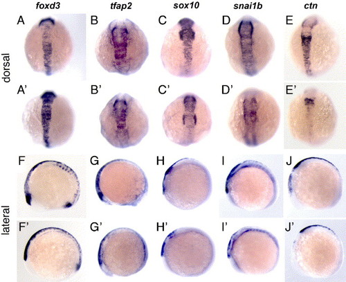
Gene expression abnormalities in premigratory neural crest populations in sym1 mutants. Dorsal (A–E, A2–E2) and lateral (F–J, F2–J2) views of neural crest expression of foxd3, tfap2a, sox10, snai1b and ctn in 10-somite stage wild-type (A–J) and sym1 mutant (A2–J2) embryos. The neural crest expression of sox10, snai1b and ctn is reduced in sym1 mutants. tfap2a expression is indistinguishable between mutant and wild-type embryos. Neural crest expression of foxd3 appears to be increased in sym1 mutants compared to wild-type embryos. EXPRESSION / LABELING:
|
|
Normal development of Rohon–Beard sensory neurons, neural plate and neural plate border in sym1 mutants. Dorsal views of wild-type (A, C, E) and sym1 (B, D, F) mutant embryos. (A, B) huC expression in 3-somite stage embryos show Rohon–Beard sensory neurons (arrowheads) in wild-type and sym1 mutant embryos. (C–F) The ectodermal neural plate and neural plate border domains, are respectively revealed by sox2 expression at the 6-somite stage (C, D) and msxb expression at the 14-somite stage (E, F) in wild-type and sym1 mutant embryos. EXPRESSION / LABELING:
|
|
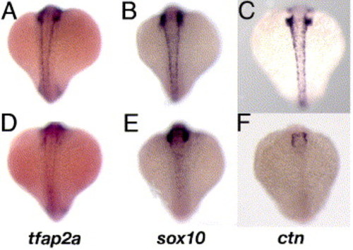
Neural crest gene expression in 18-somite sym1 mutants. Dorsal trunk views of wild-type (A–C) and sym1 (D–F) embryos. Trunk neural crest expression of tfap2a (D), sox10 (E) and ctn (F) is strongly reduced in sym1 mutants. EXPRESSION / LABELING:
|
Reprinted from Developmental Biology, 292(1), Stewart, R.A., Arduini, B.L., Berghmans, S., George, R.E., Kanki, J.P., Henion, P.D., and Look, A.T., Zebrafish foxd3 is selectively required for neural crest specification, migration and survival, 174-188, Copyright (2006) with permission from Elsevier. Full text @ Dev. Biol.


