|
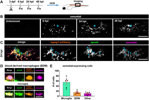
Sema4ab is expressed mainly in microglia in the spinal injury site.
(A) Experimental timeline and schematic of a zebrafish larva indicating the lateral view of the spinal injury site shown throughout the article (unless specified differently) are shown. (B) A time-course analysis of HCR-FISH indicates increased expression of sema4ab during spinal cord repair. (C) Triple-multiplexed HCR-FISH at 24 hpl shows co-localization of sema4ab, mpeg1:mCherry, and apoeb at 24 hpl (blue arrowheads). (D) High magnifications of apoeb/mpeg1:mCherry/sema4ab labeling to differentiate blood-derived macrophages (BDMs; apoeb−) from microglia (apoeb+) for cell counting. (E) Quantifications at 24 hpl show that most sema4ab-expressing cells are microglia (apoeb+/mpeg1:mCherry+/sema4ab+). Error bars show SEM. Asterisks indicate the injury site. Scale bars: 100 µm (B, C), 15 µm (D). Data files for graphs available in S2 Data.
|