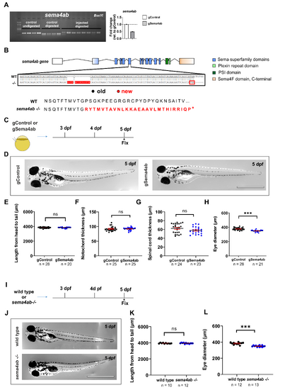
Efficient disruption of sema4ab does not lead to large changes of larval growth.
(A) RFLP and qPCR showing gRNA injection efficiency for sema4ab. For RFLP each lane represents one larva with and without digestion with the indicated restriction enzymes and with and without targeting these sites with gRNAs as indicated. Targeting the recognition sites with haCR gRNAs leads to efficient somatic mutation, as indicated by the almost complete resistance to digestion. Note a ~50% reduction in mRNA abundance detected by qRT-PCR. (B) A schematic of the sema4ab gene structure and position of the mutation in the sema4ab germline mutant is shown. Note the insertion of 14 bp in exon 6 (sequence underlaid in red) and the frameshift that predicts a stop codon (red frame and asterisk). (C) A schematic indicating the experimental design for D-H is shown. (D) Photomicrographs of gControl and gSema4ab larvae are shown at 5 dpf. (D–H) Quantifications show no differences in body length (E: gControl: 3,836 µm ± 15.65; gSema4ab: 3,838 µm ± 15.72; Unpaired t test: p = 0.9917), notochord thickness (F: gControl: 92.09 µm ± 1.527; gSema4ab: 92.20 µm ± 0.9314; Mann–Whitney U test: p = 0.3423) or spinal cord thickness (G: gControl: 62.49 µm ± 2.215; gSema4ab: 57.83 µm ± 1.677; Mann–Whitney U test: p = 0.1134) between gControl and gSema4ab larvae. Eye diameter is slightly reduced in gSema4ab larvae (H: gControl: 378.2 µm ± 3.320; gSema4ab: 349.3 µm ± 2.784; Unpaired t test: p < 0.0001). (I) A schematic indicating the experimental design for I-K is shown. (J) Photomicrographs show wild type and germline mutant larvae for sema4ab at 5 dpf. (K) No difference was observed in body length of wild type and mutant larvae (wild type: 3,941 µm ± 15.95; sema4ab −/−: 3,940 µm ± 17.50; t test: Unpaired t test: p = 0.6855). (L) Eye diameter of mutants is slightly decreased (wild type: 383.3 µm ± 3.788; sema4ab −/−: 349.9 µm ± 2.56; Unpaired t test: p < 0.0001). Each dot for qRT-PCR represents a pool of 50 larvae. β-actin was used as housekeeping gene. Raw data for qRT-PCR can be found in S5 and S6 Data. Error bars show SEM. Scale bars = 1 mm. Data files for graphs available in S3 Data. Original gels with no adjustments can be found in S1 Raw Images.
|
