FIGURE

Fig 2

ID
ZDB-FIG-260629-129
Publication
Docampo-Seara et al., 2026 - The microglia-derived protein sema4ab attenuates regenerative neurogenesis after spinal cord injury in zebrafish
Other Figures
All Figure Page
Back to All Figure Page
Fig 2

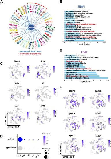
scRNA-seq indicates changes in cell-cell interactions between microglia and other cell types after sema4ab ablation.
(A) CellChat analysis for MM#1 shows interactions with all cell types and a general decrease in these interactions (blue lines) when sema4ab is disrupted. Interactions of MM#1 with ERGs (black box), fibroblasts (red curved line) and MM#2 (pink box) are highlighted. (B) KEGG analysis of the main cluster that expresses sema4ab (MM#1) shows changes in immune activation pathways (highlighted in red). (C) Feature plots show enrichment of pro-inflammatory cytokines and inflammatory markers (il1b, tnfa, il6, saa, il11b) in microglia (apoeb, circle). (D) Dot plot showing reduction of expression of pro-inflammatory cytokines and inflammatory markers after sema4ab ablation. (E) KEGG analysis of the signaling pathways most affected by sema4ab disruption in fibroblast-like cell cluster FB#2 is shown. tgfb signaling and ECM interaction-related terms are highlighted in red. (F) Feature plots showing enrichment of anti-inflammatory cytokines (tgfb1a, tgfb1b, tgfb2, tgfb3) in fibroblasts (pdgfra, pdgfrb; circled).

Expression Data

Expression Detail
Antibody Labeling
Phenotype Data

Phenotype Detail
Acknowledgments
This image is the copyrighted work of the attributed author or publisher, and ZFIN has permission only to display this image to its users. Additional permissions should be obtained from the applicable author or publisher of the image. Full text @ PLoS Biol.